A color-changing material inspired by squid skin
DOI: 10.1063/pt.ucxx.gysc

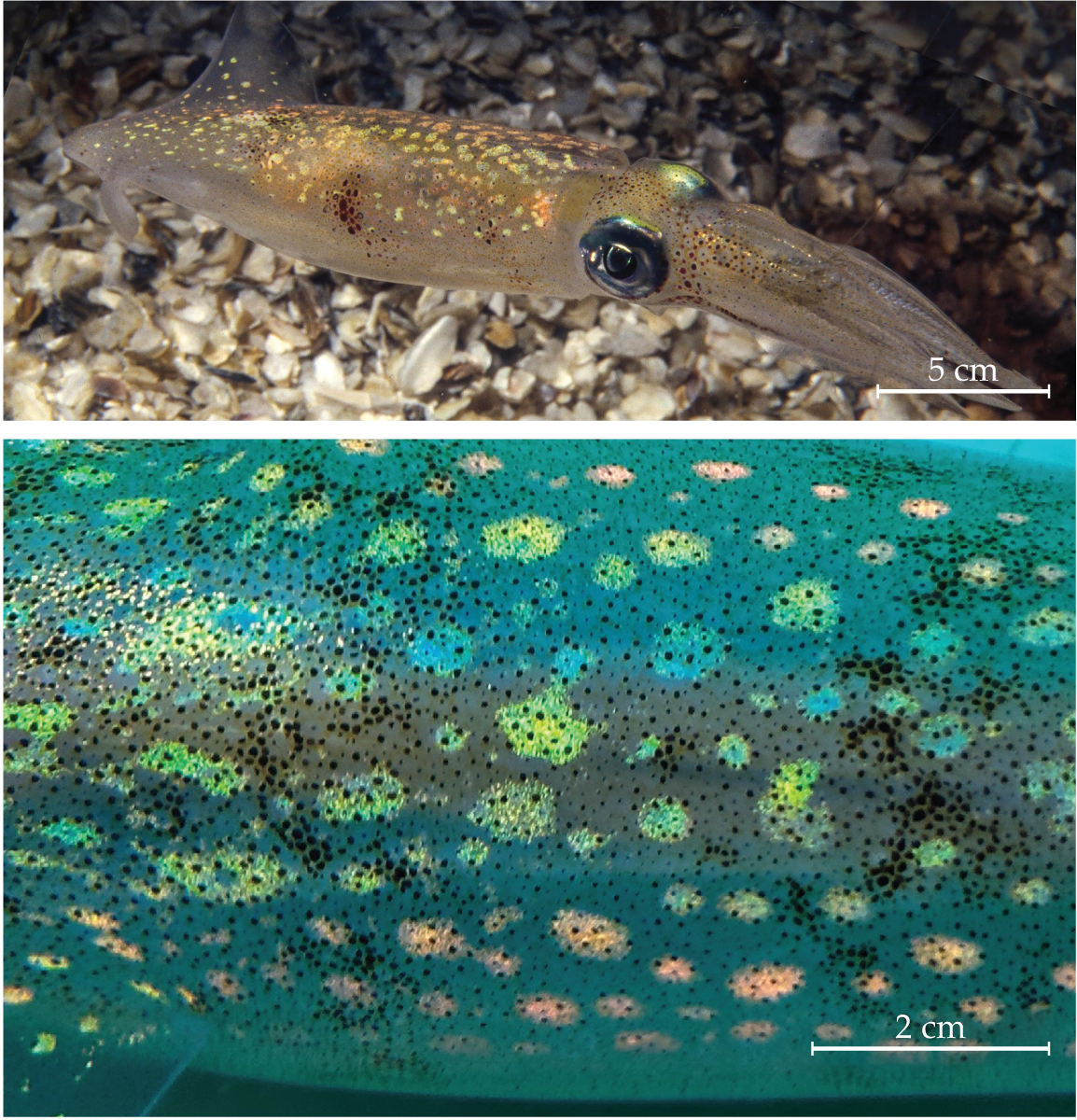
Figure 1.

Specialized light-refracting cells in the skin of the longfin inshore squid (Doryteuthis pealeii) enable it to iridesce, change color, and even become transparent. The apparent color of the skin depends on the spacing of specialized platelike proteins, known as reflectins, that produce a sinusoidal refractive index gradient. The iridescent blue, green, and orange splotches on the skin are packed with reflectins. The dark brown dots that cover the skin are chromatophores—pigmented cells that can expand and contract to change the skin’s appearance. (Images adapted from G. Bogdanov et al., Science 388, 1389, 2025 .)
Squids are well known for their camouflaging abilities. Specialized cells in their skin enable them to change color or become transparent at a moment’s notice. Now researchers led by Alon Gorodetsky at the University of California, Irvine, have made a synthetic material that uses the same tricks as squid skin to iridesce, change color, and become transparent when stretched or chemically altered. The researchers collected detailed cellular-scale observations of squid skin with a method known as holotomography—3D laser measurement of a material’s refractive index. What they found provided the key to replicating the effect.
The color-changing abilities of some squids and other cephalopods are the result of a variety of specialized cells or cell groups (see the Quick Study by Lydia Mäthger, Physics Today, December 2022, page 62 ). Cells known as chromatophores contain pigments that are revealed when the cells expand and hidden when the cells contract. Other cells, called iridophores, contain stacks of light-scattering platelike proteins, known as reflectins, surrounded by cytoplasm. Constructive interference of the light transmitted and scattered by the reflectins produces iridescence, as shown in figure
It was already understood that iridophores’ color effects were produced by the distinct refractive indices of the reflectins and cytoplasm. Gorodetsky and colleagues set out to measure those properties in 3D at the cellular level. With the help of researchers at the Marine Biological Laboratory in Woods Hole, Massachusetts, PhD student and co–first author of the study Georgii Bogdanov collected holotomography measurements of iridophores from dozens of squids. He found that most iridophores are packed with winding columns of reflectin platelets. Along the length of those columns, the refractive index varies as a sine function, with reflectins producing high values and cytoplasm producing low values.
Figure 2.

A synthetic material mimics the refractive index gradients in specialized squid-skin cells. Changing the view angle, stretching the material, or submerging it in water or other fluids can induce color changes and transparency. (Images adapted from G. Bogdanov et al., Science 388, 1389, 2025 .)
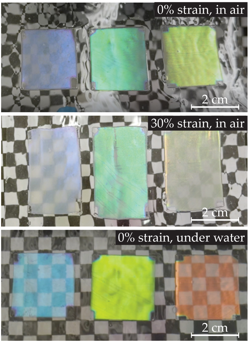
Computer simulations show that the refractive index profile’s shape is key to the color effects—triangular and rectangular profiles don’t produce the color purity or transparency that the sinusoidal profile does. The view angle, platelet periodicity, and surrounding medium also change the perceived color and transparency produced by the reflectin stacks. Using those insights, Aleksandra Anna Strzelecka, a PhD student and co–first author of the study, set about building a material that could replicate the color-changing effects.
The researchers used vapor deposition to build nanocolumns of metal oxides and varied the deposition angle to produce the targeted sinusoidal refractive index profile. The nanocolumns were then attached to a polymer to provide flexibility. The result was a material that changes color depending on what angle it is viewed from, how much it is stretched, and what fluid it is submerged in, as shown in figure
This article was originally published online on 24 July 2025.
More about the authors
Laura Fattaruso, lfattaruso@aip.org


