Targeted Ultrasound Mediates the Delivery of Therapeutic Genes to Heart Muscle
DOI: 10.1063/1.2169431
If you start a program of vigorous exercise, your muscles will sprout new capillaries. Behind that performance-enhancing response lies an elaborate interplay of genes and proteins. Conceivably, the same molecular machinery could be switched on to make new blood vessels in a diseased heart. Then, doctors could induce heart muscle to grow arteries that bypass life-threatening blockages, sparing their patients the sliced flesh and cracked bones of open-heart surgery.
That’s one of the hopes for cardio-vascular gene therapy. If realized, it would work roughly as follows. Identify a protein whose presence causes blood vessels to form. Produce and package strands of DNA that contain the gene for making the protein. Deliver the DNA to heart muscle.
Of those steps, the last is the most challenging. The heart lies within the rib cage and behind the lungs. With each beat, it fluctuates in volume by about 25%. Directly injecting DNA into that well-defended moving target is dangerous. Delivering the DNA in specially modified viruses—a standard technique in experimental gene therapy—is also dangerous. Even if they don’t cause infection, modified viruses still provoke the body’s immune system, and they don’t necessarily end up where the DNA is needed.
In the late 1990s, physicians and physicists hit on the idea of using ultrasound contrast agents to deliver DNA. The contrast agents consist of micron-sized gas-filled bubbles encased in shells made of lipids, proteins, or sugars. When exposed to ultrasound, the bubbles pulsate resonantly, reflecting the ultrasound and boosting the detected signal.
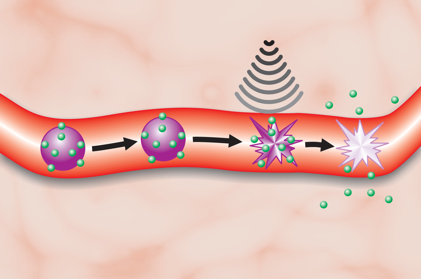
If the ultrasound is intense enough, the bubbles can burst with sufficient force to breach the membranes of nearby cells. And if the bubbles are coated with DNA, their destruction releases the DNA, enabling it to enter the cells through the holes forced open by the burst. The accompanying figure depicts the process.
Early experiments proved that ultrasound-targeted microbubble destruction (UTMD) does indeed breach cells. 1 Followup work successfully transferred genes that encode readily detectable markers, such as luciferase, a protein responsible for the glowing abdomen of the male firefly.
Now, a team led by Paul Grayburn of Baylor University Medical Center in Dallas, Texas, and Ralph Shohet of the University of Texas Southwestern Medical School, also in Dallas, has demonstrated that UTMD can deliver genes that stimulate the growth of new blood vessels in rat heart. 2 UTMD, they hope, could lead to minimally invasive treatments for heart disease, diabetes, and other diseases.
Making DNA-coated microbubbles is surprisingly simple. The Dallas researchers shake DNA, in the form of short circular strands called plasmids, in a saline solution with the other ingredients: lipids for the shell and a hydrocarbon for the gas. DNA-coated bubbles then self-assemble and can be sorted by size.
The bubbles should be small enough that they can pass through the lungs’ tiny blood vessels. In that case, the bubbles circulate throughout the body but burst only when they pass through the ultrasound beam at the organ of interest. A bubble diameter of several microns is typical.
For their experiment, Grayburn, Shohet, and their collaborators used DNA that encodes a protein called vascular endothelial growth factor. Ordinarily, muscle and other cells secrete VEGF when they don’t receive enough oxygen. When VEGF sticks to the lining of blood vessels, it triggers the production of more vessels and restores the oxygen supply.
Proving that UTMD-delivered DNA causes the formation of new blood vessels required several careful controls. Rats treated with ultrasound and bubbles could conceivably express VEGF, even if the bubbles lack DNA. To eliminate that possibility, the Dallas researchers used DNA that encodes human VEGF, rather than rat VEGF. Rat cells respond readily to both. Finding the human protein in rats after UTMD proved the corresponding gene was successfully transferred.
The Dallas researchers divided their rats into four groups. Each group received ultrasound, which was directed at the heart in four bursts using a commercially available transducer. Only one group of rats received DNA-coated bubbles. The rest, in three control groups, were injected with uncoated bubbles, free DNA, or a saline solution.
After intervals of 5, 10, and 30 days, rats within each group were killed and their heart tissue examined. Only the group injected with DNA-coated bubbles showed a significant increase in the density of blood vessels.
Interestingly, the increase was transient: 13% after 5 days, 27% after 10 days, none after 30 days. The use of plasmid DNA, which isn’t stably incorporated into a cell’s genome, could account for the transience.
Improvements
Even though UTMD evidently mediates gene transfer, exactly how it does so is unclear. The bubbles collapse at speeds approaching 1 km/s, which is to fast to film. Modeling the bubbles is just as hard. The bubbles’ collapse is extremely nonlinear and, when it occurs in narrow blood vessels, is strongly influenced by vessel walls. Computer models suggest that if the collapse is asymmetric, narrow jets shoot out and could serve as the battering rams. Mechanically sensitive transmembrane proteins might also play a role.
There’s another mystery. Genetic transcription takes place in the cell nucleus. Somehow, UTMD enables DNA to penetrate not only the cell membrane, but also the nuclear membrane.
A deeper understanding of UTMD would help solve one of the many problems that beset gene therapy however it’s mediated: low efficiency. Most of the administered DNA never triggers protein production. Still, despite the lack of understanding, there are plausible paths forward. The ultrasound’s spectrum, intensity, and time structure can all be tweaked, as can the bubble composition and the choice of gene.
If perfected, UTMD offers a way of delivering therapeutic genes noninvasively to hard-to-reach organs. Grayburn and his collaborators are already testing the technique on another such organ, the pancreas.

Gas-filled microbubbles (purple) covered with DNA (green) pass harmlessly and uneventfully through blood vessels until they are exposed to ultrasound. Then, the bubbles burst, causing not only the release of the DNA but also the opening of holes in the cells that line the vessel.
(Courtesy of Ralph Shohet.)
References
1. R. J. Price et al. Circulation 98, 1264 (1998) https://doi.org/10.1161/01.CIR.98.13.1264 .
2. G. Korpanty et al. Gene Therapy 12, 1305 (2005) https://doi.org/10.1038/sj.gt.3302532 .


