A laser selectively kicks carbon out of a foil
DOI: 10.1063/PT.3.4916
An intense laser can ionize matter and accelerate the resulting ions into a beam. Creating and accelerating ions in a single compact system offers an advantage over the bulky conventional combination of a separate ion source and a cyclotron or synchrotron. Such laser-driven ion beams could be useful in various applications, including radiotherapy, that benefit from short high-flux pulses of ions. 1 (See the article by Jerimy Polf and Katia Parodi, Physics Today, October 2015, page 28 .)
But controlling what ions end up in the beam can be difficult in laser-acceleration systems. The problem is that the target materials inevitably contain contaminants, primarily hydrogen. When the laser hits, protons from the ionized hydrogen dominate the beam and prevent heavier ions from accelerating.
Although proton therapy is currently the most widespread ion radiotherapy, a treatment that employs larger ions, such as carbon, interests medical researchers because the ions’ extra mass means they do more damage to a cancerous cell’s DNA as they pass through. A reliable source of carbon-ion beams that is small enough to fit in a typical laboratory would facilitate more research on potential carbon-ion therapies. (See Physics Today, June 2015, page 24 .)
Now Marco Borghesi of Queen’s University Belfast in Northern Ireland and his colleagues have developed a laser method to selectively excite heavier ion species. 2 Their complementary simulations demonstrate how the shape of their laser pulse preferentially moves protons out of the way so that carbon ions can be accelerated.
A new regime
Lasers can accelerate ions out of a material in a few ways. The most common method is known as target normal sheath acceleration (TNSA). In it, the laser ionizes surface atoms and produces free electrons that work their way through the material and escape out the back. As they leave, they create a strong electric field that ionizes and accelerates atoms on the back surface. Because TNSA acts on the surface atoms, it disproportionately accelerates contaminants, which sit primarily on the surface and are difficult to avoid or remove.
Radiation-pressure acceleration, on the other hand, acts on the bulk of the material. With sufficiently high intensity, the light pulse applies pressure directly on the electrons and ions and pushes them out of the material. Such acceleration happens in two regimes that depend on the target thickness. For thicker samples, the hole-boring regime prevails: Light pressure dents the target, and the excited ions must push their way through the rest of the material.
For thinner samples, tens of nanometers thick, the acceleration enters what’s known as the light-sail regime, in which the ions escape directly. Although that acceleration is more efficient than the hole-boring regime, thinner targets are difficult to work with. For example, the laser profile needs to be temporally smooth because any sudden spikes in intensity that precede the main pulse can prematurely destroy a thin target.
Hit the target
The Borghesi group has worked on laser techniques for accelerating ions since 2005. In 2013 the group joined A-SAIL (Advanced Strategies for Accelerating Ions with Lasers), a consortium of several UK universities established to explore whether those techniques could be used for medical applications. Several years later Borghesi and his A-SAIL collaborators were the first to publish convincing results that they were reaching the light-sail acceleration regime for amorphous carbon foil targets. 3 The researchers found that carbon ions were accelerated efficiently, but the beam still contained a mix of ion species, with the protons, because of their lighter masses, at higher energies per nucleon. Although the target had about a sixth as many protons as carbon ions, at high energies the ion beam had around 500 times as many protons as carbon ions.
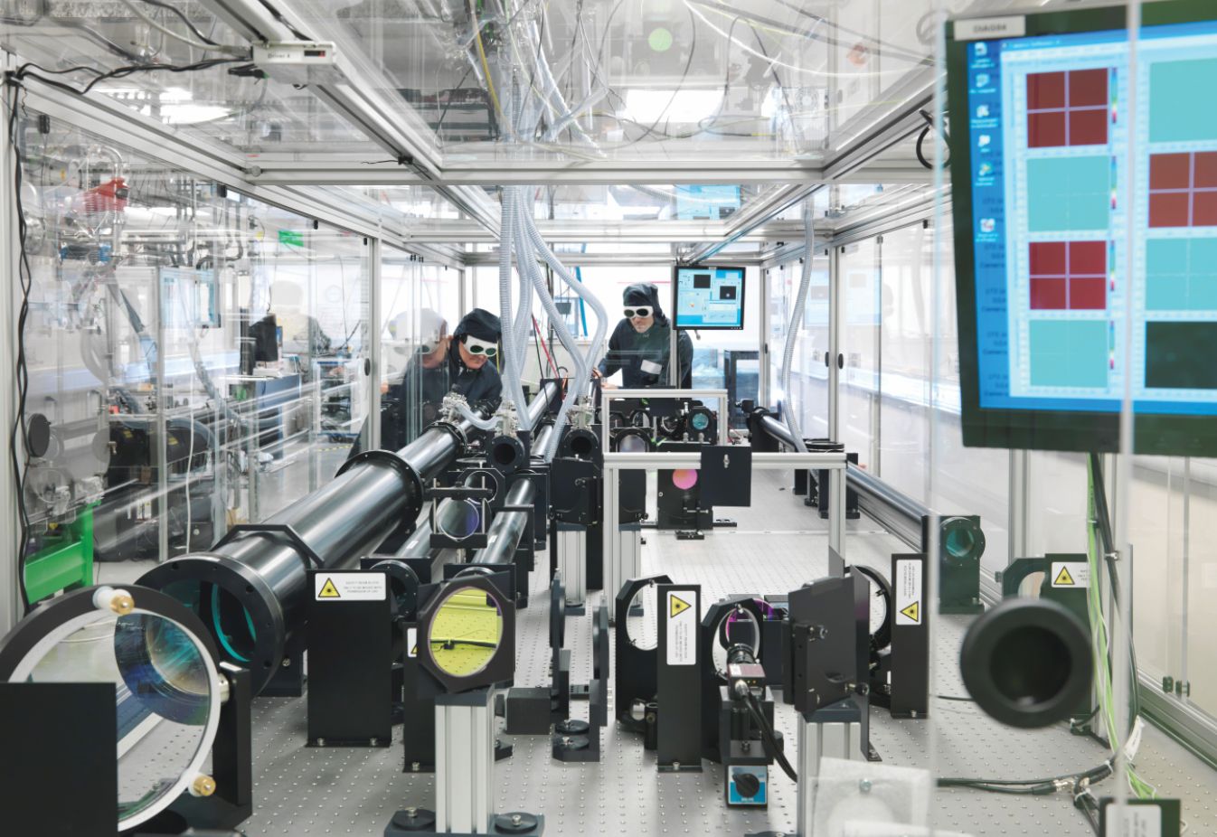
Borghesi and his then graduate student Aodhan McIlvenny decided to try again with more targets at a wider range of thicknesses than they used with the previous work. They performed the study at the Central Laser Facility, shown in figure
Figure 1.

The Gemini laser at the Central Laser Facility, part of the UK’s Rutherford Appleton Laboratory, provided pulses for a recent experiment on laser-driven ion acceleration. Here researchers check the alignment before they send the laser beam to the target chamber. The shape of the pulses’ temporal profiles resulted in the preferential acceleration of carbon ions over proton contaminants that usually dominate ion beams. The short, high-flux burst of carbon ions could be useful for research into more efficient radiotherapy treatments. (Courtesy of the Central Laser Facility, STFC.)
The researchers were looking for the optimal target thickness. Intuitively, the thinner the sample is, the more energy each ion will get from the laser. But to experience radiation acceleration, the material must reflect the laser beam.
Reflection happens only when the material exceeds a critical density at which it becomes opaque to the laser frequency. For highly intense laser pulses, the electrons in the plasma move at relativistic speeds, and their effective masses thus increase. The critical density consequently increases, so a target that was once opaque becomes transparent. That so-called relativistically induced transparency sets in at lower laser intensities in thin samples than in thicker ones.
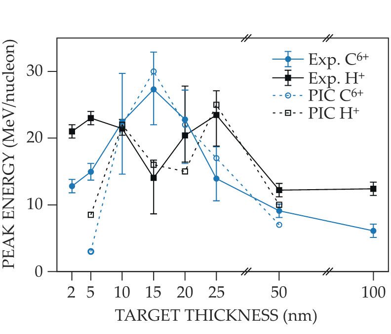
In the new study, McIlvenny measured the maximum energy of the ejected carbon ions as a function of target thickness and found that the peak occurred in a 15-nm-thick target, as shown by the solid blue line in figure
Figure 2.

Carbon ions (C6+) reach a peak energy per nucleon in experiments (solid blue line) and in particle-in-cell (PIC) simulations (dashed blue line) when laser accelerated out of a target that’s 15 nm thick. That optimal thickness strikes a balance of thin enough that a laser can easily push ions out of the material but not so thin that the laser has trouble interacting with it. Surprisingly, proton contaminants (H+) in the material have a local energy minimum for the same target thickness (solid and dashed black lines). In previous experiments, the protons always had higher energies per nucleon than carbon ions did. (Adapted from ref.
In theory
To understand and verify the atypical behavior, McIlvenny performed particle-in-cell simulations of the carbon and proton energies as a function of target thickness and laser intensity for an idealized pulse and for one matching the experimental pulse. In the case of an idealized Gaussian temporal profile, the carbon ions’ and protons’ energy trends roughly matched, although the protons’ energies were 30% higher.
The realistic laser profile, on the other hand, had a shoulder before the peak intensity. That shoulder resulted from the two plasma mirrors that the researchers used to smooth the laser profile and increase its intensity contrast. A plasma mirror is a polished surface that when excited by a high-intensity laser forms a plasma, which acts as a highly reflective active optical element. For the leading edge of the laser pulse, the plasma mirrors suppress the intensity until the laser is powerful enough to make them reflective, as happened a few picoseconds before the peak intensity.
The pulse shape creates three stages in the laser–foil interaction. First, the low-intensity shoulder causes the target to expand radially out from the center of the laser spot. Protons, with their lighter masses, race away from the central region faster than carbon. Next, the rising intensity causes the material in the central region to contract, but few protons remain in the region by that point. Finally, the peak arrives and accelerates ions, which are primarily carbon, in the central region. Protons are accelerated afterward by a less-efficient mechanism similar to TNSA. The simulations showed that the same species-selective acceleration should be possible with a pair of pulses.
In practice
Other methods exist to reduce the contaminants in ion beams, including running an electric current through the target before irradiation and ablating the material first with another laser. Most of the methods, however, are complicated to implement and too destructive for the sorts of thin samples needed to access the efficient light-sail acceleration regime.
In the future, Borghesi and his colleagues’ technique could be used for studying the effects of irradiation in cell cultures and other biological samples to help develop more efficient radiotherapy treatments. To get meaningful and reproducible results that can be compared with those from conventional sources, researchers need a well-defined ion species with a well-defined energy distribution. The light-sail acceleration setup offers those benefits in a system compact enough for a typical lab. The technique also accesses unique regimes of cellular irradiation—in particular, shorter bursts of ions than are possible with a conventional accelerator. Shorter doses offer the advantage of less damage to nearby healthy cells.
Borghesi and his collaborators are already exploring those research topics. Right now, the ion beam is useful for planar cell cultures, but moving to three-dimensional in vitro or even in vivo studies will require higher-energy carbon ions. The team members are exploring ideas for how to boost the energy: employing recently developed high-power lasers, gaining better control of the temporal and spatial shapes of the laser pulse, and finding ways to delay a thin target from becoming relativistically transparent. They are also exploring techniques for focusing the ion beam, which would offer even higher doses.
References
1. J. Schreiber, P. R. Bolton, K. Parodi, Rev. Sci. Instrum. 87, 071101 (2016). https://doi.org/10.1063/1.4959198
2. A. McIlvenny et al., Phys. Rev. Lett. 127, 194801 (2021). https://doi.org/10.1103/PhysRevLett.127.194801
3. C. Scullion et al., Phys. Rev. Lett. 119, 054801 (2017). https://doi.org/10.1103/PhysRevLett.119.054801


