Covalent organic frameworks aid in mass-spectrometry imaging


(Images courtesy of the Zhibin Yin Laboratory, Institute of Advanced Science Facilities, Shenzhen, China.)
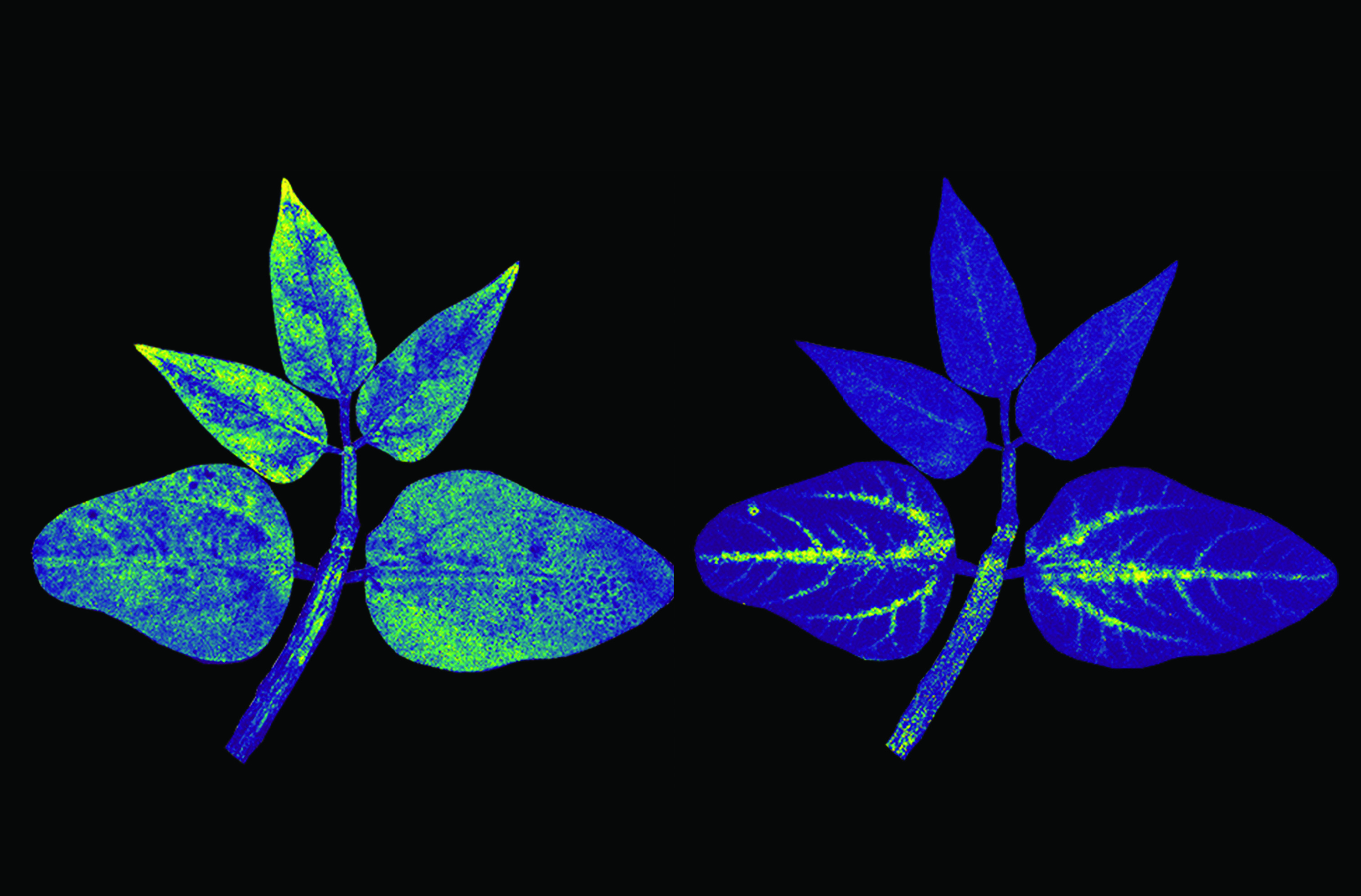
These mass-spectrometry images of a cowpea plant reveal two embedded compounds: dinotefuran (left), a pollutant with which the plant was irrigated, and histidine (right), an amino acid naturally produced by the plant as part of its metabolic process. The light regions in both images show the molecules of dinotefuran and histidine, respectively, as determined via their distinct mass-to-charge ratios. The dinotefuran has spread throughout the plant, whereas the histidine molecules have accumulated in the vascular areas of the larger, mature leaves.
To determine mass-to-charge ratios in a mass spectrometer, the sample needs to be ionized. That’s a trivial task for simple substances, but historically, researchers have had trouble with large biological molecules, which tend to fragment when ionized. That began to change in the 1980s and 1990s with the development of such techniques as surface-assisted laser desorption/ionization (SALDI), in which the sample is deposited onto a substrate that absorbs energy from the ionizing laser. But the nanomaterials used to fabricate the substrate can be laborious to create and difficult to consistently mass produce.
To generate mass-spectrometry images of an entire cowpea plant, a feat that had not been done before, a team led by Zhibin Yin at the Institute of Advanced Science Facilities in Shenzhen, China, developed a new type of SALDI substrate that adds nanofilms made of covalent organic frameworks to a commonly used substrate base made with titanium oxide nanotubes. Because the new substrate can be easily produced and integrated into the methods typically used to prepare biological specimens, the researchers hope that it will become standard in the laboratory and enable the imaging of even larger organisms.
Reference
1. Y. Xu et al., “Interface-engineered plasmonic covalent organic framework nanofilms on TiO2 nanotubes for universal mass spectrometry imaging ,” Sci. Adv. 12, eadx8264 (2026).


