The brain is big science
DOI: 10.1063/PT.3.2207

Two major initiatives seek breakthroughs in understanding the human brain. Broadly, the US emphasis is on developing new experimental tools, while a European project focuses on modeling and computing.
This fall the agencies participating in the US Brain Research through Advancing Innovative Neurotechnologies (BRAIN) Initiative, unveiled on 2 April by President Obama, began requesting proposals. The public–private BRAIN Initiative has $110 million in federal money for fiscal year 2014: $40 million from the National Institutes of Health, $20 million from NSF, and $50 million from the Defense Advanced Research Projects Agency (DARPA). The total budget and time scale for the project are “not really spelled out,” says Thomas Insel, director of the NIH’s National Institute of Mental Health. “But the president made clear that this is the beginning and that it will require a longer and larger commitment.”
On the private side, collaboration and investments totaling around $100 million a year will come from the Kavli Foundation, the Howard Hughes Medical Institute (HHMI), the Salk Institute for Biological Studies, and the Allen Institute for Brain Science, which is already sharing the petabytes of data arising from the brain project it launched last year.
In January the European Commission announced the selection of the Human Brain Project (HBP) as one of two future and emerging technologies flagship programs, each with expected funding of €1 billion ($1.3 billion) from the commission and member states over 10 years (see Physics Today, July 2011, page 25 , and page 22 of this issue). Some 200 groups from 80 institutions are participating so far, and about 20% of the initial proposals will support people not yet involved in the HBP.
Tool revolution
“We don’t have a single unifying theory for how the brain works,” says Miyoung Chun, the Kavli Foundation’s executive vice president of science programs. “We know the microscale of single neurons, and we know how large patches—the macroscale—of brain light up in fMRI [functional magnetic resonance imaging], but we are lacking [knowledge about] how the brain works at the mesoscale level. If we can have systematic and comprehensive tool development, we should be able to go far.”
The directions for BRAIN research are still being defined. DARPA announced in October that it will focus on deep brain stimulation. And an interim advisory report to NIH lays out nine areas to pursue, including generating a census of cell types, creating structural maps, and linking neuronal activity to behavior. It’s not easy to be specific about methods that don’t yet exist, but some of today’s cutting-edge research may offer clues of what’s to come.
“The best way to understand the human brain is to study simpler brains,” says Loren Looger, a protein engineer at the HHMI Janelia Farm Research Campus in Virginia. He makes optogenetic probes that encode an animal’s genes to produce fluorescent indicators of neural activity. “The hunger for these tools is great,” he says. The only brain that is fully mapped at the structural level is that of the ubiquitous lab worm Caenorhabditis elegans, he notes. “That nervous system has 302 neurons, and even there, many mysteries remain.” The human brain has around 100 billion neurons, each with thousands of synapses. “In the vast gulf separating worms and humans are mice and fruit flies—both of which are standard research models,” says Looger.
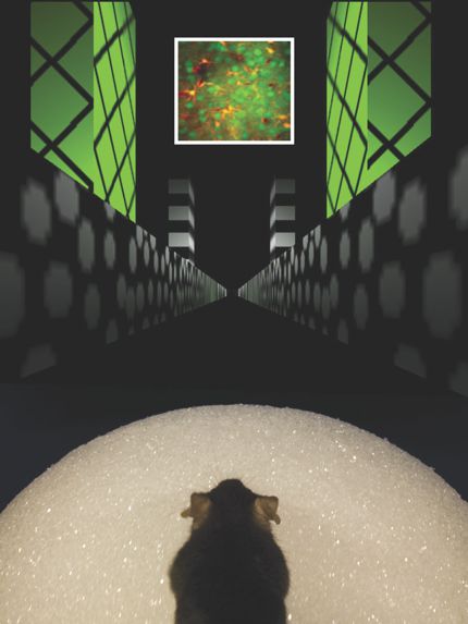
Biophysicist David Tank at Princeton University couples probes such as Looger’s with two-photon microscopy to watch neurons fire as mice navigate virtual mazes (see the photo at left). The mice, Tank says, “know it’s not a real maze, but all the evidence we have suggests they are engaging the same cognitive systems as when they navigate in the real world.”

Reading a mouse’s mind as it navigates virtual reality. The inset at top shows a population of neurons imaged with two-photon microscopy as a mouse, its head held in place, ran atop the Styrofoam ball.
Forrest Collman, Daniel Dombeck, Chris Harvey, and David Tank
Cornell University’s Chris Xu has built a three-photon microscope to push the limits of depth imaging. He looks at the brains of live mice, usually with some skull removed. “I think I can go to 2 mm in depth,” he says. That goes through layers of gray and white matter to reach subcortical neurons. The white matter is especially challenging, he explains, because with its fatty myelin protection of nerve fibers, it strongly scatters light. From an optical point of view, says Xu, “it’s clear what we need: noninvasive imaging over a large volume in awake animals with high spatial resolution—down to a single cell or better.”
Another approach to imaging deeper into tissue is adaptive optics. In telescopes, adaptive optics involves deforming light-collecting mirrors to correct for wavefront aberrations introduced by the atmosphere. In microscopy, says Janelia Farm physicist Na Ji, the trick is to shape the wavefront of the excitation light “to cancel out the brain-induced aberrations and form a sharp focus deep inside the brain, so that neurons can be imaged at subcellular resolution.”
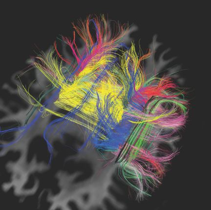
Van Wedeen, a radiologist and neuroscientist at Harvard University and the Martinos Center for Biomedical Imaging, wanted to see if he could map the wiring of the brain with MRI by tracking how water diffuses along signal-carrying fibers in white matter (see the image above). “As you increase the resolution, instead of becoming more complicated, the images get simpler and prettier. It turns out the fibers of the brain to a surprising extent form a [regular] three-dimensional grid. I am not referring to the small scale of axons. What we are seeing is the structure of large pathways from one area to another,” he says.

Diffusion magnetic resonance imaging noninvasively tracks water molecules in a living adult human brain, revealing that pathways of brain wiring form a grid pattern. Each false-color line represents about 105 axons, or nerve fibers.
V. Wedeen and L. Wald, Martinos Center for Biomedical Imaging
The past few years have seen a revolution in tools to study the mouse brain, says NIH’s Insel. “We now have spectacular opportunities to do precise monitoring and large-scale studies on the dynamics of the mouse brain. We have almost none of that for the human brain.” What’s needed, he says, is new tools. “They don’t come from biology, they come from engineering, materials science, physics.” Insel notes that NIH’s $40 million for the BRAIN Initiative may seem like a drop in the bucket compared with the institute’s $5.5 billion annual investment in neuroscience. But, he says, “development for tools and technologies doesn’t fare well in our grants system. So this makes a difference by saying, ‘Let’s create enabling technologies.’”
Smart simulations
By the time it was selected as a flagship, Europe’s HBP was well defined. The emphasis is on computing, simulations, and aggregating data. The project is best known for its goal of simulating a human brain.
An integrated approach is needed because so far people “are running into the brain recording anything and everything” and producing “an exponentially growing amount of fragmented data,” says project architect Henry Markram, a neuroscientist at the Swiss Federal Institute of Technology (EPFL) in Lausanne, Switzerland. Most of the brain’s organization, he says, “could be predicted from fundamental rules. We are after those rules.”
The project “is a huge convergence between information technology and life sciences,” says HBP spokesman Richard Walker of EPFL. “We would like to construct multilevel atlases of the brain—from large-scale fibers, to individual cell types, down to gene expression. What we are trying to do is exploit the fact that there is a huge amount of repetition in the brain.” Humans are different from mice, but there are similarities—basic synapses are similar, he says. “We want to leverage that.” Early brain simulations are to be available for users to test hypotheses in 2016.
The HBP researchers “want to build a brain from the basic elements—nerve cells, glial cells, connectivity,” says Thomas Lippert, director of supercomputing at Germany’s Jülich Research Centre, “then approximate the electrical and chemical networks.” At the same time, new knowledge and constraints about how the brain functions will be incorporated into models, he says. “Eventually, you could remove or add connections and simulate diseases or tumors.”
Lippert’s group boasts the most detailed 3D human brain model to date. It has voxels 20 microns to a side and was created from MRI scans of thin slices of a real brain. Alan Evans of the McConnell Brain Imaging Center at McGill University in Montreal, Canada, which collaborated on the model, says, “It’s a high-resolution scaffold that allows you to assemble data from other scales—genetic, metabolic, neurochemical, . . .”
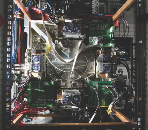
Karlheinz Meier, a physicist at Heidelberg University and one of three HBP directors, builds neuromorphic computers (see the photo at right). “Rather than simulating circuits from differential equations, we use microelectronics to build physical copies of brain cells, connections, and synapses.” Analog circuits use about 100 picojoules for a synaptic signal, compared with 1 J for a supercomputer simulation and 1 fJ for a biological system, says Meier. And whereas supercomputers are up to 1000 times slower than live systems, neuromorphic computers are actually faster—thanks to their even tinier components. “A day’s [brain] activity can be compressed into 10 seconds,” he says. “This leads to really interesting experiments, for example, on synaptic plasticity and learning.” The silicon neurons in the Heidelberg lab’s neuromorphic brain each have 20 variable parameters. “Some neurons fire once, some regularly, some in bursts. Some adapt to stimulus and then adjust. We can emulate all of those patterns.”

This neuromorphic computing system in Heidelberg mimics 200 000 neurons and 50 million synapses on a silicon wafer located behind the central octagonal aluminum plate. Field-programmable gate arrays configure the network and send and read input and output data.
Heidelberg University
Seductive and polarizing
The HBP has stirred controversy among scientists, with many saying it promises too much. They ask, How can one simulate the brain when so little is known about it? A neuroscientist who requested anonymity says that Markram “makes gorgeous images and hypnotizes the crowd. When I heard him talk, part of me was saying, ‘I will follow him to the end of Earth.’ The other part of my brain was saying, ‘That is nonsense.’ It was extraordinary. I was surprised how powerful the seduction was.”
Supporters of the HBP dismiss the criticism. McGill’s Evans blames the media and funding agencies for the hype around the European project. “You cannot get the public’s and politicians’ attention unless you make a big pitch,” he says. “It doesn’t matter if the project does not reach its ultimate goal of simulating the entire human brain. Much good science will come out of it.” As to simulations being premature, Lippert says, “Simulations constrain experimental research. Not simulating would exclude an important approach to science. The promise is only to increase the understanding of the brain.”
For his part, Meier says, “If everyone is for a project, it’s probably not that interesting. If everyone is against it, it’s probably wrong. But if it’s polarizing, that is a good sign.”
Big data, big implications
Scientists in the US and Europe see the two big brain projects as complementary. The techniques developed in the BRAIN Initiative will be used to gather new data, which, in turn, will feed into the simulations of the HBP.
But the masses of data “that will come gushing out from all those labs” are a “potential showstopper,” says Terry Sejnowski, a neuroscientist at the Salk Institute. “The current state of data storage and analysis will have to be amped up.” At the same time, he says, “there will be a major effort to make the data public—as is the norm in astronomy and genomics but not in neuroscience.”
An overarching goal is to understand the brain in both health and disease and eventually to help treat such disorders as depression, epilepsy, Alzheimer’s and Parkinson’s diseases, and autism. “I have two agendas,” says Harvard’s Wedeen. “One is to understand how the brain works. And we are urgently in need of a revolution in mental health.”
Beyond leading to treatments for disease, the brain projects “will galvanize industry,” says the Kavli Foundation’s Chun, pointing to robotics as one area likely to benefit from research on the brain. “It’s only three pounds of mushy material that, using little energy, can do unbelievable things,” she says. “Imagine if we can unlock its secrets.”
More about the authors
Toni Feder, tfeder@aip.org


