Physicists invited to apply their insights to cancer
DOI: 10.1063/1.3431325

Can physicists help get cancer research out of a rut? That’s what the National Cancer Institute is betting on with the roughly $150 million it’s spending over five years on a network of 12 centers, each a multi-institutional, multidisciplinary collaboration led by a physical scientist. “We’re taking cancer, turning it on its side, giving it to a new group of people, and seeing what happens when we combine what we already know with what they come up with,” says Larry Nagahara, NCI program director for the Physical Sciences-Oncology Centers (PS-OCs). The centers got started last fall.
Cancer research hasn’t seen a major paradigm shift in 30 or 40 years, according to William Grady, who studies signal deregulation and epigenetic modifications in gastrointestinal cancer at the Fred Hutchison Cancer Research Center in Seattle. “Most advances involve revisions and refinements. We do not have medical treatments that can cure [most cancers] despite decades of effort.”
The idea behind the PS-OCs is to bring cancer research fresh perspectives, such as relating disease to the physical properties of cells. “What’s new is thinking not only in terms of the chemical concentrations of signaling proteins, but also in terms of their spatial organization within the cell,” says physicist Jan Liphardt, principal investigator of the PS-OC led by the University of California, Berkeley. Another example, he says, is that tumors are firmer than surrounding tissue. “What has not been known is whether this stiffening is a bystander, or if mechanical changes are intimately involved in cancer progression.” To find out, researchers in Liphardt’s PS-OC injected mouse breasts with cells engineered to crosslink collagen and then added cancer cells. They found that cancer grew faster in breasts that had been preconditioned to be stiff.
One project at the MIT-based PS-OC involves “measuring the instantaneous growth rate of single cells at known points in the cell cycle,” says Scott Manalis, whose team developed a resonator that can weigh single cells to femtogram resolution. “We can measure mass, density, and charge. There are many interesting clinical questions we can ask with this,” he says, “such as, Can these physical properties of cells tell you how patients will respond to therapeutics?”
“The thing about cancer,” says physicist Paul Davies, who heads up the PS-OC based at Arizona State University, “is that you are never going to solve the problem by details. You cannot micromanage. It always outmaneuvers you.” The cell, Davies says, is so “stupendously complex we will never figure out on the level of individual interactions what to do. But we may not need to. If physical features turn out to trigger cancer, we don’t need to know all the gory details.”
The PS-OCs are not “another exercise in asking physicists to give oncologists another beam weapon,” says Davies. “Physics has been used in cancer treatment—x rays, proton bombardment. What [NCI is] after here are insights from physicists to be brought to bear on cancer.”
Says Princeton University physicist Robert Austin, “I am not convinced cancer is a disease. It’s an inevitable part of evolution. We will never get rid of it. We need to learn to live with it. That is a different approach.” Researchers have a “miserable track record of looking at cancer the wrong way,” he adds. The collaborations he is involved with through the PS-OC he heads are a “shotgun marriage,” he says. “It’s stressful, but it’s time to make the leap. Maybe we can make an impact on cancer fundamentals. To do that, I will have to dance with these other people.”
The PS-OCs are special, Liphardt says, “because of how rare it is to have funding to do something really new, and just try things.” The locations, principal investigators, and other information relating to the 12 centers can be found online at http://physics.cancer.gov. .

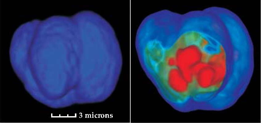
Robust methods for early cancer detection may be developed from tomographic imaging of single cells. These images show the nuclear surface (left) and a slice from the reconstructed isotropic three-dimensional image (right); DNA density increases from green to red. The images are formed with computed tomography, but whereas clinical CTs use an x-ray source and detector, here the source was white light and the detector a microscope equipped with a CCD camera.
(Courtesy of Vivek Nandakumar, Laimonas Kelbauskas, Roger Johnson, Deirdre Meldrum, Center for Ecogenomics, Biodesign Institute, Tempe, AZ.)
More about the authors
Toni Feder, American Center for Physics, One Physics Ellipse, College Park, Maryland 20740-3842, US . tfeder@aip.org


