Novel Medical Imaging Method Shows Promise
DOI: 10.1063/1.2117810
Scientists from Philips Research in Hamburg, Germany, are developing a new method for peering inside patients. Magnetic particle imaging, as the method is called, resembles magnetic resonance imaging in its use of magnetic fields to manipulate spins. But whereas MRI flips hydrogen nuclei, MPI flips the electronic spins of ferromagnetic particles.
The naturally occurring ferromagnetic particles in the human body, such as the iron atoms in hemoglobin, are too small to produce a detectable magnetization. Using an MPI-based medical scanner, if one were ever built, would entail introducing nanoscale tracers into the body through a syringe, catheter, or other device.
But the absence of a natural background means the injected tracers would provide the sole signal. Potentially, MPI offers exquisite sensitivity.
Whether MPI will lead to a practical scanner is unclear. So far, the method’s developers, Bernhard Gleich and Jürgen Weizenecker, have looked at modestly sized test objects. Even so, MPI has already matched MRI in spatial resolution and, Gleich and Weizenecker hope, should soon exceed it in sensitivity. 1
Harmonic magnetization
What makes MPI possible is the characteristic S-shaped magnetization curve of a ferromagnet. As the accompanying
Ordinarily, an induction coil placed around the magnetized tracers would record the fluctuating signal from both the weak magnetization and the much stronger field. But if a frequency filter excludes the fundamental, only the harmonics from the magnetized tracers contribute to the detected signal.
By themselves, the harmonics reveal the general presence of magnetized tracers. To locate the tracers, Gleich and Weizenecker exploit another part of the magnetization curve. Adding a constant field to the periodically varying field can shift the tracers’ magnetization into the saturation region and, as a result, quench both the fundamental and the harmonics.
The trick of quenching the harmonics helps to locate the tracers because it’s possible to engineer a field-free point within the constant field. Only tracers in the volume around the point remain unsaturated and free to emit harmonics. By steering the point through the sample like a probe, one can map the tracer distribution.
The spatial resolution of MPI depends on how steeply the magnetic field rises from zero at the field-free point to the value H k required to produce detectable harmonics. If the field gradient is G, the expected resolution is roughly 2H k /G.
The value of H k depends on the volume of a tracer particle: The bigger the particles, the lower the field required to overcome thermal fluctuations and align the magnetization. Paradoxically, big tracers give high resolution.
In their proof-of-principle experiment, Gleich and Weizenecker used an FDA-approved ferromagnetic tracer called Resovist. According to the bottle’s label, Resovist’s characteristic particle size is 4 nm, which, at the gradients routinely available in human-sized scanners, would yield a useless resolution of 10 cm. Fortunately, as Gleich and Weizenecker discovered, a few percent of Resovist particles are more than 30 nm in diameter. At 0.3 mm, the resolution produced by the larger particles compares favorably with commercial MRI.
Magnetization of ferromagnetic nanoparticles

Magnetic particle imaging depends on the particles’ S-shaped magnetization curve, like the one shown here. At low values of applied magnetic field H, the magnetization M is linear. At high values of H, M saturates. In the intermediate regime (indicated in gray), M rolls over and can be expressed as an expansion in odd powers of H:
where a 1, a 3, and so on are constants. Now, if the applied field varies periodically, say, if H = H 0sin ωt, then, thanks to trigonometric identities such as 4sin3 θ = 3sin θ – sin 3θ,
Here, f 1, f 3, and so on are polynomial functions of H 0. The harmonics at 3ω and 5ω can be detected by filtering out the fundamental at ω.
Another key consideration in MPI’s performance is how one steers the field-free point. Stepping the point through the sample—or, equivalently, stepping the sample through the point—is the most straightforward and robust method, but it’s slow because the scan has to stop for each microliter-sized volume element.
A faster method is to sweep the point back and forth at high frequency. In effect, the motion smears the signal over neighboring volume elements. That’s not necessarily a problem. The signal can be reconstructed through standard signal inversion. However, to avoid stifling the reconstruction with large errors, the point’s trajectory must be highly stable.
Sweeping has another benefit. If done fast enough, the motion can induce harmonics by itself without the need for a periodically varying field. The frequency required is of order 10 kHz, which would be challenging and potentially hazardous to effect mechanically. Instead, Gleich and Weizenecker use three electromagnets whose fields can rapidly displace the field-free point in the x, y, and z directions.
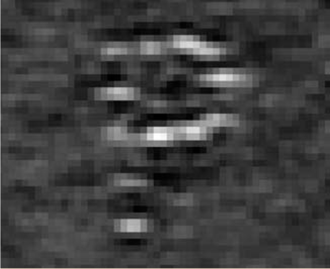
The figure below shows the result of imaging a test object, a plastic tray drilled with holes 1 mm deep and 0.5 mm wide that contain Resovist. Gleich and Weizenecker used a hybrid scanning method to make the image. A robot moved the field-free point in the xy plane, while in the z direction the point was moved electromagnetically.

Thirteen holes, each 0.5 mm in diameter, form the letter P, which can be seen in this test image. The field of view is about 1 cm square.
(Adapted from ref. 1.)
MPI’s eventual commercialization will depend on several improvements. Optimizing the tracer size is one. Based on a straightforward comparison with MRI, Gleich and Weizenecker anticipate that MPI, thanks to its almost-zero background, should be 100 times more sensitive than MRI.
Scanning is a bigger obstacle. The electromagnets Gleich and Weizenecker currently use can shift the field-free point by no more than 6 mm on each sweep. Short sweeps mean long scanning times. The 9.4 × 9.4 mm2 test object took 1 minute to scan. Using stronger electromagnets and higher sweep frequencies will shorten the overall time needed to map a body-part-sized object.
What might MPI be used for? Following the flow of blood and nutrients through capillaries requires a combination of sensitivity and resolution that MRI lacks and that x rays provide only with long and possibly harmful exposure times.
References
1. B. Gleich, J. Weizenecker, Nature 435, 1214 (2005) https://doi.org/10.1038/nature03808 .


