Neutron Diffraction Overcomes Flux Limits to Resolve a Large Protein Structure
DOI: 10.1063/1.1634521
On paper, thermal neutrons seem ideal for probing the structure of crystallized biomolecules. At a few tenths of a nanometer, the de Broglie wavelength of thermal neutrons matches molecular bond lengths. And thermal neutrons interact strongly enough with matter to pick up structural information but weakly enough to pass through crystalline samples.
Another desirable property of thermal neutrons has to do with hydrogen. X rays, the most commonly used structural probe, interact with the atomic electric field; the more electrons an atom has, the stronger the interaction. With just one electron per atom, hydrogen, the most abundant element in biomolecules, is all but invisible in x-ray diffraction patterns.
Neutrons interact with nuclear spins. Though predictable, the strength of the interaction oscillates with apparent randomness across the periodic table. Hydrogen, it turns out, scatters neutrons with about the same strength as potassium.
Scattering strength also varies from isotope to isotope. Conveniently for crystallographers, one of the strongest differences is between hydrogen and deuterium. Substituting deuterium for hydrogen at a specific chemical site reveals the site’s spatial location through a simple comparison of diffraction patterns.
Knowing where hydrogen atoms are is invaluable for understanding proteins, especially enzymes. All proteases (the enzymes that cut proteins into pieces) work by exchanging hydrogen atoms, as do many isomerases (the enzymes that restructure proteins). Half, if not more, of the reactions catalyzed by proteases and isomerases would be easier to understand if certain hydrogen atoms could be located.
Unfortunately, despite its advantages, neutron diffraction has one big drawback: It’s difficult to produce neutrons in large numbers. Whereas third-generation synchrotrons flood samples with 1019 x-ray photons per second per square centimeter, a reactor source, such as the one at the Institut Laue–Langevin in Grenoble, France, emits about 1015 neutrons per second per square centimeter.
Because of the relatively low intensity of neutron beams, it takes crystallographers far longer to collect a neutron diffraction pattern than an x-ray diffraction pattern. And more copies of the molecule have to be present in the neutron beam. The practical upshot is that crystals have to be large and the crystallized molecules small. In the first-ever use of neutrons to determine the structure of a protein, Benno Schoenborn spent a whole year, 1968, gathering reflections from a 25-mm3 crystal of myoglobin, a 17-kilodalton transport protein. 1
Since Schoenborn’s pioneering experiment, the flux of neutron sources has increased, as have the sensitivity of neutron detectors and the effectiveness of analysis techniques. Nevertheless, until recently it was impractical to obtain clear neutron diffraction patterns from the many large and interesting proteins that weigh 50 kDa or more.
That limit now looks set to disappear. In a proof-of-principle experiment, a team from Los Alamos and Oak Ridge national laboratories and the Fox Chase Cancer Center in Philadelphia has succeeded in using neutrons to determine the 1.8-Å structure of xylose isomerase, a 160-kDa, industrially important enzyme. 2
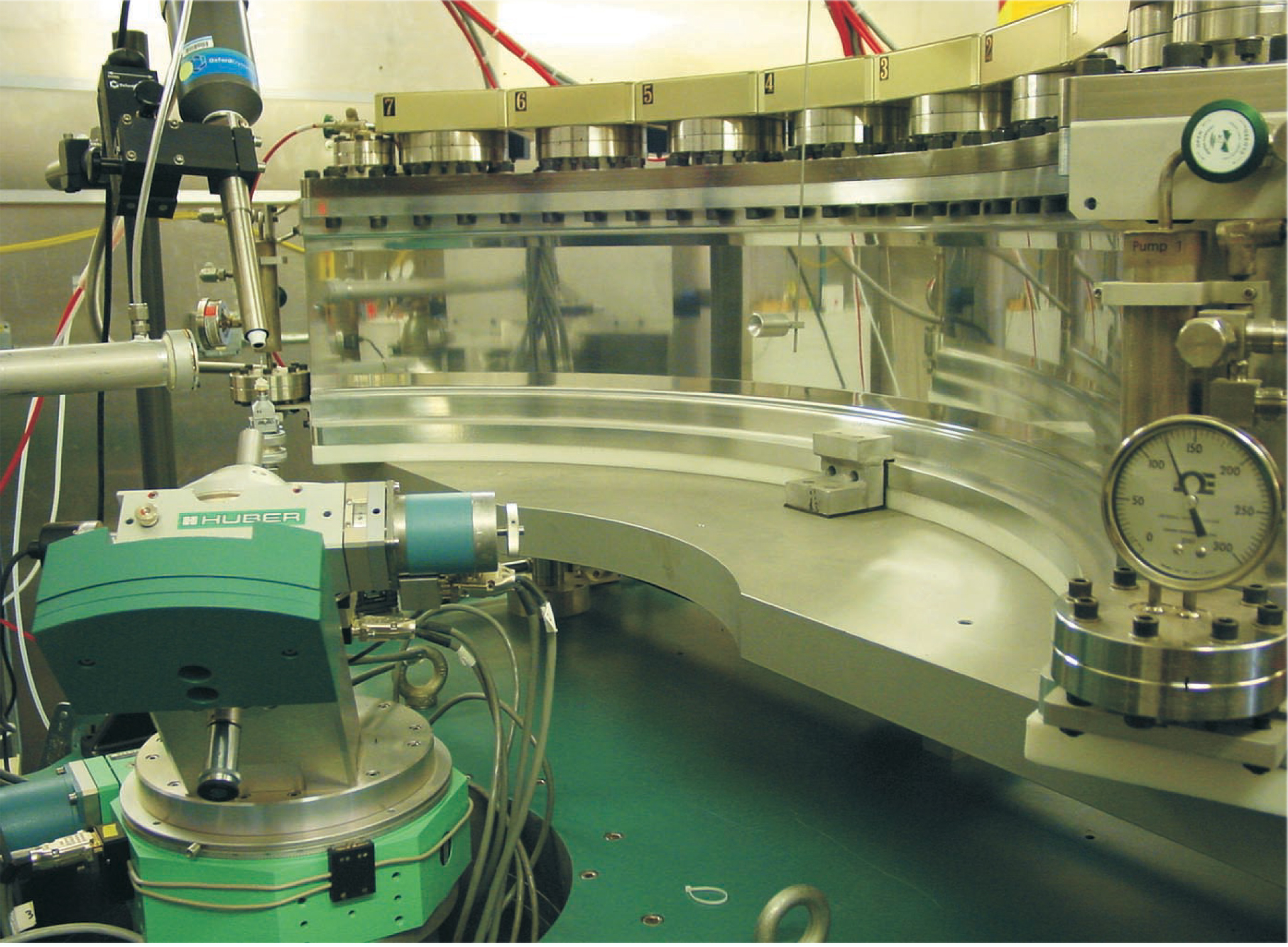
The team, led by Gerry Bunick of Oak Ridge and Leif Hanson of the University of Tennessee, could tackle such a large protein thanks to the spallation source at the Los Alamos Neutron Science Center (LANSCE) and the detectors at the center’s Protein Crystallography Station. Commissioned two years ago and shown in figure 1, PCS is beginning to produce its first results. It’s also paving the way for a similar station at the Spallation Neutron Source, which is scheduled to start generating neutrons at Oak Ridge in 2006.

Figure 1. The Protein Crystallography Station at the Los Alamos Neutron Science Center. The sample is held by the rotating stage at the bottom left. Eight detectors are arrayed about 1 meter away from the sample to collect the scattered neutrons.
(Courtesy of Gerry Bunick.)
All the available neutrons
There are two types of thermal neutron source: nuclear reactors, which produce neutrons during the fission of enriched uranium, and spallation sources, which produce neutrons when accelerated protons smash into a metal target.
Neutrons emerge from both sources with a broad distribution of wavelengths. That feature is important because the quantity one measures in a diffraction experiment, the Bragg angle, yields the separation of scattering planes only when one knows the wavelength of the scattered particles.
In reactor sources, the neutron wavelength is selected using a monochromator, which filters out all but a narrow range of wavelengths. Recent improvements to analysis techniques have made it possible to widen the wavelength window. Still, 95% of the neutrons end up unused.
Because spallation neutrons are produced by an accelerator, they can be timed and their wavelengths determined. By using time-of-flight techniques in combination with detectors that tag every event, LANSCE’s PCS gathers wavelength-resolved data from all the available neutrons. Consequently, even though LANSCE produces fewer neutrons than a big reactor, it can bring more neutrons to bear on a sample. The ability to resolve data in wavelength also leads to higher peak-to-background ratios and lower backgrounds.
Candy bars and soda pop
Bunick and Hanson chose to work on xylose isomerase in part because of its role in the food industry. Nowadays, food manufacturers use high-fructose corn syrup instead of sucrose to sweeten candy bars, soda pop, and other commercial comestibles. Two enzymes are essential for producing the syrup: amylase, which breaks down corn starches into simpler sugars, and xylose isomerase, which converts one of those simpler sugars, glucose, into fructose. By weight, fructose is two times sweeter than sucrose and nearly three times sweeter than glucose.
Glucose and fructose share the chemical formula C6H12O11, but differ structurally: At the first and second carbons, glucose has H–C=O and H–C–OH, whereas fructose has H2–C–OH and C=O. To convert glucose to fructose, xylose isomerase somehow helps hydrogen atoms move from the second carbon to the first. If they knew the exact mechanism, food industry scientists could try to make the conversion more efficient—or at least see whether their effort would be fruitless.
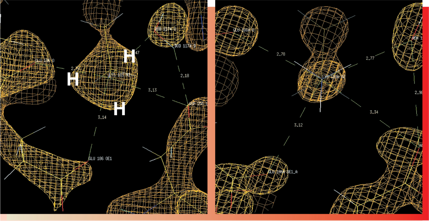
Even with their new, neutron-derived structure in hand, along with a higher-resolution structure previously derived from x rays, Bunick, Hanson, and their collaborators haven’t yet been able to nail down exactly how xylose isomerase moves the hydrogen. But they have a good idea about where to look. Figure 2 gives a flavor of the kind of data they’ve been taking.

Figure 2. The terminal nitrogen of lysine-183, a key amino acid in xylose isomerase, is the central feature of both the neutron density map on the left and the electron density map derived from x rays on the right. Unlike the electron map, the neutron map reveals the three hydrogen atoms around the terminal nitrogen of lysine-183.
(Courtesy of Gerry Bunick.)
Xylose isomerase is one of several proteins that contain doubly charged metal ions—in fact, it has two. A magnesium ion occupies one site; a magnesium, cobalt, or manganese ion occupies the other. The ions are vital to the enzyme’s function: If you take them away, the enzyme can neither bind to the glucose substrate nor catalyze the conversion.
Being strongly positive, metal ions attract the oxygen atoms in water molecules. Negatively charged parts of the enzyme attract water’s hydrogen atoms. Subjected to such attractions, a water molecule will depolarize. And if the depolarized water molecule is close enough, it can help the hydrogen atoms on the second carbon atom of glucose hop over to the first carbon.
Solving the mechanism will involve tracking the movement of site-hopping hydrogens with respect to the two metal ions, the amino acids of the enzyme, and nearby water molecules. To do that, the team plans to investigate the structure of the enzyme-substrate complex.
References
1. B. P. Schoenborn, Nature 224, 143 (1969) https://doi.org/10.1038/224143a0 .
2. B. L. Hanson et al., Trans. Am. Crystallogr. Assoc. (in press).


