Nanoparticles Locate and Flag the Blood Vessels That Nourish Tumors
DOI: 10.1063/1.1628992
Cancer kills its victims when the tumor burden in their bodies reaches about 1013 cells. Before then, when a tumor comprises about 1010 cells and is the size of a sugar cube, a physician can usually find it in an x ray or MRI scan. At that stage, if treated promptly, a victim may survive.
Too often, however, cancer lurks unnoticed until it’s past curing. Lack of resolution isn’t what frustrates early detection. Clinical MRI and x-ray machines can resolve features a few hundred micrometers across. Rather, the difficulty lies in identifying a small blob of cells whose combined malignancy is too weak to signal where to look and whose appearance in an MRI or x-ray image is too similar to the surrounding, healthy tissue.
But at the molecular level, cancer cells look and behave quite differently from healthy cells. They reproduce faster and, as they do so, manufacture certain molecules in far greater numbers.
Among a cancer cell’s molecular output are transmembrane proteins called integrins. Integrins take part in angiogenesis, the process by which tumors build networks of blood vessels that tap into the host’s blood supply. Without siphoned nutrients, a tumor can’t grow beyond a few tens of cells.
Like some other proteins, integrins can be disabled when antibodies of just the right shape lock on to them. In 1998, Dorothy Sipkins and her collaborators at Stanford University and the Scripps Research Institute exploited integrin’s role in angiogenesis and the specificity of its antibodies to create a new method for flagging tumors. 1
Sipkins’s idea was to coat liposomes (artificial, cell-like capsules of fluid) with integrin antibodies and MRI contrast agents. Thanks to the antibodies, the liposomes would attach to the sites of angiogenesis. The contrast agents would act as beacons, boosting and sharpening the MRI signal.
At 300–350 nm across, the coated liposomes qualified—metrically speaking—as nanoparticles. To test them, Sipkins implanted small tumors into the hind legs of rabbits. After giving the tumors time to form new blood vessels, she injected the liposomes into the rabbits’ bloodstreams. Twenty-four hours later, the angiogenesis sites showed up in MRI scans.
Now, a team led by Greg Lanza and Sam Wickline of Washington University in St. Louis (WUSTL) has extended the technique. 2 Like the Stanford—Scripps team, the WUSTL researchers coat nanoparticles with integrin antibodies and MRI contrast agents, but instead of liposomes, they use an emulsion of nanoscale droplets, which are easier to make and carry more contrast agents. Testing their nanoparticles on rabbits, they see significant MRI enhancement after just two hours.
Lanza and Wickline intend to register the nanoparticles with the US Food and Drug Administration as what the agency calls an investigational new drug. Clinical trials should begin in 2006.
Oil, water, and lipids
Making nanoparticles the WUSTL way is a bit like making salad dressing. Instead of olive oil and balsamic vinegar, Lanza and Wickline use water and an octane derivative called perfluorooctylbromide (PFOB). And instead of a whisk, they use a microfluidizer, a device that forces mixtures through a fine sieve to create an emulsion of nanoscale droplets, shown schematically in figure 1.

Figure 1. Targeted nanoparticles, shown in schematic cross section, contain liquid perfluorooctylbromide encased in a functional lipid skin.
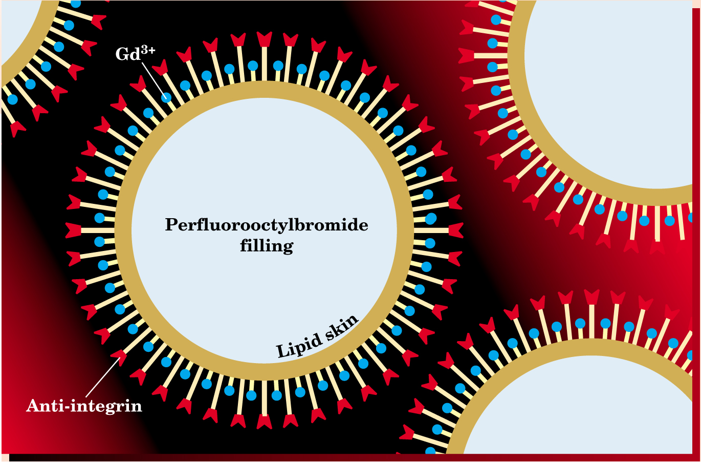
The immiscibility of PFOB and water also provides a way to coat the droplets. When mixed with PFOB and water, lipids seek the interface between the two liquids. At the interface, the lipids’ hydrophilic heads point into the water, while their hydrophobic tails point into the PFOB. Coating a droplet with functional groups is simply a matter of binding the groups to the lipids’ hydrophilic ends, a task that can be done before mixing and emulsification.
For the antibody, Lanza and Wick-line use a synthetic peptide. For the MRI contrast agent, they use gadolinium ions. Thanks to their seven unpaired electrons, Gd3+ ions are extremely paramagnetic and, following an MRI pulse, can help protons in their vicinity regain equilibrium faster than distant protons. This difference in recovery time (T 1 relaxation time to cognoscenti) shows up as enhanced contrast. The 90 000 Gd3+ ions that cover a typical WUSTL nanoparticle speed up T 1 relaxation rates by a factor of a million.
The WUSTL nanoparticles are about 250 nm in diameter. If the particles were much larger, lungs would trap them. If the particles were smaller, they couldn’t carry enough contrast agents to enhance images.
Twelve rabbitsz
During angiogenesis, proliferating cancer cells break through the walls of the host’s blood vessels and create holes. The blood that feeds the growing tumor leaks through the holes and through the newly forming vessels.
Because of this leakiness, any nanoparticles, whether studded with sticky antibodies or not, can make their way out of the bloodstream and pool temporarily around the tumor. To verify that their nanoparticles adhere to the sites of angiogenesis, Lanza and Wickline divided their experimental subjects—12 rabbits—into three groups of four.
The targeted group received integrin-targeted, gadolinium-covered nanoparticles. The control group received nontargeted gadolinium-covered nanoparticles. The competitive blockade group received a dose of targeted, uncovered particles two hours before imaging began. That group then received a dose of targeted, covered nanoparticles.
All 12 rabbits were implanted with a tumor, each about 2 × 2 × 2 mm3 in size. The WUSTL researchers then waited for 12 days before injecting the rabbits with the nanoparticles. That’s enough time for a tumor to initiate angiogenesis, but not enough time for it to grow significantly. If physicians could catch such small tumors in humans, the chance of a complete cure would be high.
After a dose of anesthetic, each rabbit was placed in a 1.5-tesla MRI chamber and imaged six times: 30 minutes before dosage, immediately after, then 30, 60, 90, and 120 minutes after. To quantify any image enhancement, the team used the predosage image as a baseline.
At first, the targeted and control groups showed similar enhancement, which the WUSTL team attributes to the leakiness of the blood vessels around the tumors. But by two hours, as more and more nanoparticles stuck to the tumors, the targeted group had moved ahead to exhibit an average enhancement of 125%.
The competitive blockade group had the lowest enhancement. As Lanza and Wickline anticipated, the first wave of targeted, uncovered particles stuck to the sites of angiogenesis and partially blocked access for the second wave of targeted covered particles.
Under the microscope
Figure 2 shows a typical MRI image from one of the rabbits in the targeted group. In this example, the patches of enhanced contrast appear on the edges of the tumor. In other examples, patches appear close to, but not on, the tumor, as if the tumor were in the process of establishing a vascular bridge.

Figure 2. Nuclear magnetic resonance image of a tumor implanted in the hind leg of a rabbit. The yellow patches in this 9 × 10 mm2 image indicate regions of enhanced NMR emission and correspond to the sites of angiogenesis.
(Adapted from ref. 2.)
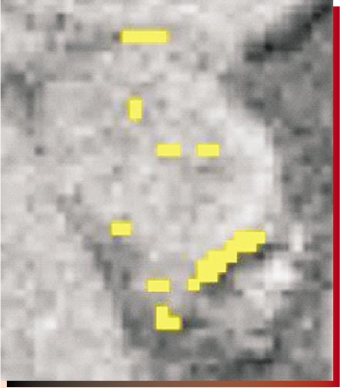
To confirm that the patches correspond to angiogenesis, the researchers cut out the tumors, then stained, sliced, and examined them under the microscope—a process called histology. The histology provided confirmation.
Proving that targeted nanoparticles can locate and stick to the sites of angiogenesis is important not only for diagnosing cancer, but also for potential therapies. In 1971, Harvard University’s Judah Folkman hypothesized that stopping angiogenesis would contain cancer. 3 Now, several angiogenesis inhibitors, among them the drugs endostatin and angiostatin, are in clinical trials. And last year, David Cheresh of Scripps and his collaborators showed that integrin-targeted nanoparticles can deliver therapeutic genes to cancer cells. 4
Testing any anticancer drug involves assessing its effect on tumors. Targeted nanoparticles could do the job without the need for a biopsy. And they don’t need MRI. Lanza points out that PFOB is twice as dense as water, which makes it an x-ray contrast agent, and has a sound speed one third that of water, which makes it an ultrasound contrast agent, too. Not bad for salad dressing.
References
1. D. A. Sipkins et al., Nat. Med. 4, 623 (1998) https://doi.org/10.1038/nm0598-623 .
2. P. M. Winter et al., Cancer Res. (in press).
3. J. Folkman, N. Engl. J. Med. 285, 1182 (1971) https://doi.org/10.1056/NEJM197108122850711 .
4. J. D. Wood et al., Science 296, 2404 (2002) https://doi.org/10.1126/science.1071599 .


