Low-dose phase-contrast mammography
DOI: 10.1063/PT.3.2136
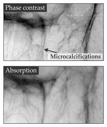
Small, treatable tumors are difficult to spot in mammograms because healthy and cancerous tissues differ little in how they absorb x rays. But absorption isn’t the only source of contrast. As x rays pass through an inhomogeneous medium, they can acquire differences in phase—even if the medium is a uniform absorber. Early attempts at phase-contrast imaging required a synchrotron or other bright, coherent source of x rays. Now Alessandro Olivo of University College London and his collaborators have built a prototype machine that performs phase-contrast mammography with a conventional x-ray tube at clinically acceptable doses. The setup works by masking the x-ray source with an array of hundreds of narrow, closely spaced holes. Each beam that emerges points at a single pixel of a flat-panel detector. The detector is also masked—such that half the x rays from each beam are prevented from reaching their designated pixel. When an object is placed between the source and the detector, the beams suffer either absorption or, thanks to a change in phase, refraction. Because of the setup’s geometry and the small angles of refraction involved, some refracted photons will still reach their pixel, but others will miss and hit the mask. Enough photons are diverted to the mask that they boost the contrast of what would otherwise be a conventional absorption image. Olivo’s team tested its setup on donated samples of cancerous breast tissue. Microcalcifications that presage cancer showed up more clearly in the phase-contrast image than in an absorption image. (A. Olivo et al., Med. Phys. 40, 090701, 2013.)


