Lampreys Rely on a Molecular Switch to Detect UV Light
DOI: 10.1063/1.1784267
Our biological clocks, like those of other vertebrates, keep time thanks to the response to sunlight of the pineal gland. Stuck underneath our cerebral hemispheres, the lentilsized gland can’t detect light directly. Rather, it receives signals from our eyes via a tortuous circuit of nerve cells. Whatever pineal photosensitivity our ancestors once enjoyed lost out to a higher evolutionary priority: boosting the computing power of the mammalian brain.
But the pineal of lower vertebrates is both closer to the top of their heads and better equipped to detect light. In lizards, for example, the pineal corresponds to an eyelike organ that lies beneath the skin and between the eyes.
Intrigued by this light-sensing ability, biologists wondered whether it might be linked to mysterious functions that humans and other mammals no longer perform. Experiments ensued and, from the late 1960s onward, revealed that the pineal glands of certain species of fish and amphibians can detect UV light.
For setting a biological clock, using UV would make sense. The ratio of UV to visible light is higher in twilight than in daylight. However, despite its potential utility, the pineal’s UV sensitivity has not been definitively linked to a biological purpose.
On the molecular front, things are a little clearer. By the early 1990s, biologists were close to identifying the photoreceptor molecules responsible for pineal photosensitivity. Evidence suggested that pineal photoreceptors share the same molecular plan as the photoreceptors that mediate vertebrate vision.
In visual photoreceptors, a bulky transmembrane protein called an opsin envelops a light-sensitive derivative of vitamin A called retinal. Free retinal absorbs maximally in the UV, but the electrostatic influence of the opsin shifts the absorption. Depending on what amino acids abut the retinal, the absorption maximum lies anywhere between the UV and the red.
At first, biologists hoped they could identify and isolate pineal photoreceptors by looking for genetic sequences that resemble those of the more familiar visual photoreceptors. That strategy proved difficult to pursue because the resemblances are not as strong as originally expected. But in 1994, Toshiyuki Okano of Tokyo University succeeded in identifying a photoreceptor cloned from chicken pineal. Other identifications of pineal photoreceptors followed: in the trout, toad, and catfish.
Now, a team led by Akihisa Terakita of Kyoto University and Satoshi Tamotsu of Nara Women’s University has identified a pineal photoreceptor in another vertebrate, the Japanese river lamprey Lethenteron japonica. 1 By using a multidisciplinary approach, the Kyoto-Nara team demonstrated that the newly discovered pineal photoreceptor is UV sensitive.
Moreover, unlike all other known vertebrate photoreceptors, the lamprey’s pineal photoreceptor is bistable. That is, it flips between two stable states in response to light of different wavelengths. Such bistability had been predicted based on physiological studies of frogs and other vertebrates, but hadn’t been seen before except in the eyes of insects.
Spectroscopy and genetics
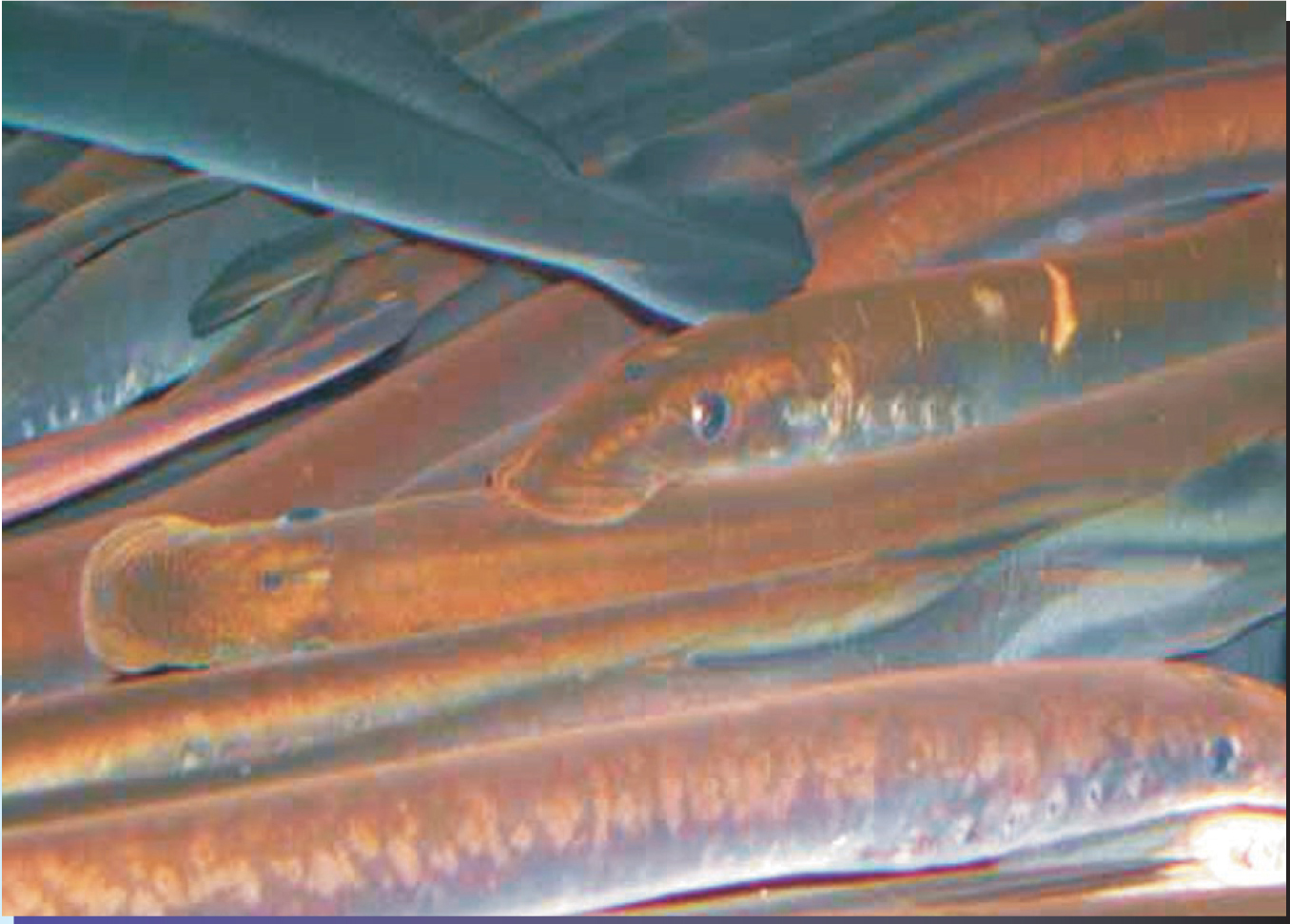
Lampreys are primitive fish. They lack jaws and bony skeletons and, as figure 1 shows, bear a superficial resemblance to eels. Terakita and Tamotsu decided to study L. japonica because its response to light had been extensively studied. In particular, Eberhardt Dodt and Hilmar Meissl found in 1982 that visible light stimulates the firing of certain pineal nerve cells, whereas UV light suppresses firing. Dodt and Meissl’s study suggested that the lamprey pineal was sensitive to the ratio of UV to visible light.

Figure 1. Japanese river lampreys grow to be about 20 cm long. For food, they latch their suckerlike mouths onto other fish to withdraw their victims’ blood.
(Courtesy of Akihisa Terakita.)
To identify the lamprey’s pineal photoreceptor, the Kyoto-Nara researchers looked for opsinlike sequences in lamprey DNA. They found five. The first two sequences resembled those of retinal G protein-coupled receptors. Although RGRs participate in the visual process, they don’t sense light. The next two sequences appeared to code visual opsins, one for red light, the other for green light. The fifth sequence was the most promising. Its closest relatives code pineal opsins found in the catfish, trout, and toad. Armed with the sequence, the researchers pressed bacteria into making numerous copies of the putative pineal opsin.
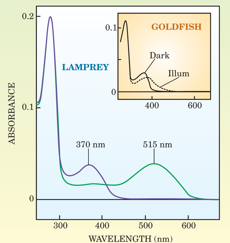
To work as a photoreceptor, an opsin needs a retinal molecule. Fortunately, opsins and retinal molecules readily pair up in solution. Kyoto’s Mitsumasa Koyanagi illuminated samples of the reconstituted photoreceptor and measured its absorbance. UV light, he found, is absorbed maximally at 370 nm and converts the photoreceptor into a state whose absorbance peaks in the green at 515 nm. In the dark, this photoexcited state proved long-lived, but illuminating the sample with orange light snapped the photoreceptor back into its original UV-absorbing state. Figure 2 shows the spectra.

Figure 2. Absorbtion spectra of the lamprey’s pineal photoreceptors (main panel) and the UV photoreceptors in the goldfish’s eye (insert). Before UV illumination, the absorbance of the lamprey’s photoreceptors (purple) peaks in the near UV at 370 nm. After illumination, the absorbance shifts to the green at 515 nm (green). By contrast, the goldfish’s UV photoreceptors lack a second absorption peak after illumination. The strong peak at 280 nm belongs to amino acids that don’t-participate in photoreception.
(Adapted from ref. 1.)
The lamprey, like other fish, has a two-part pineal composed of the pineal proper at the top and the parapineal below. To see if the UV-absorbing photoreceptor really is expressed in the pineal and, if it is, to determine its distribution, the Kyoto-Nara researchers created RNA probes for both the pineal opsin and for one of the two visual opsins they’d found in the lamprey DNA. Released into the pineal and parapineal, the molecular probes latch onto their respective opsins and stain them dark blue.
The purely visible photoreceptors turned up in the lower parts of the pineal and parapineal, whereas the UV photoreceptors turned up in the upper parts. Evidently, the pineal and parapineal have at least two distinct light-sensing regions.
Photoreceptors convert light to an electrical current that, by flowing across the cell membrane, changes the membrane’s electrical potential. To verify that the UV photoreceptors behave in the same way, Emi Kawano of Nara Women’s University inserted tiny electrodes into those pineal cells identified by the RNA probes as containing the UV photoreceptors. Illuminating the cells with UV caused the membrane potential to shoot up, which is consistent with previous studies, including Dodt and Meissl’s.
Goldfish eyes
L. japonica can’t see in the UV, but certain other fish can. Terakita decided to compare the properties of the lamprey’s pineal photoreceptors with those that mediate UV vision in the goldfish.
He and his coworkers isolated the genetic sequence of the goldfish UV opsin and reconstituted the corresponding photoreceptors, just as they had for the lamprey’s UV photoreceptor. Subjecting the goldfish photoreceptors to the same spectroscopic tests revealed a different response to UV light. The goldfish’s absorbance, like the lamprey’s, exhibits a maximum in the UV. But after exposure to UV light, no amount of light at any wavelength can restore the UV-absorbing state.
This behavior is not a surprise. When it absorbs light, retinal undergoes cis–trans isomerization. In the lamprey’s pineal photoreceptors, the retinal stays put. But in the goldfish’s visual photoreceptors, the isomerization triggers the retinal’s disassociation from the opsin. Special proteins shepherd the released retinal to another cell type, whose job it is to reverse the isomerization. Proteins then return the refreshed retinal.
This rather baroque mechanism evolved to serve the eye’s primary function: vision. Can the bistability of the lamprey’s pineal photoreceptor shed light on the pineal’s function? The spectral state of a bistable photoreceptor—and hence its effect on the cell membrane—depends on the ratio of UV to visible light. Because water absorbs and scatters UV more strongly than visible light, Terakita speculates that the lamprey could use the ratio to sense its depth beneath the surface.
References
1. M. Koyanagi et al., Proc. Natl. Acad. Sci. USA 101, 6687 (2004).https://doi.org/10.1073/pnas.0400819101


