Intense X-Shaped Pulses of Light Propagate Without Spreading in Water and Other Dispersive Media
DOI: 10.1063/1.1825261
It seems inevitable. If you send a short, narrow pulse of light through a dispersive medium, the light’s component colors, traveling at different speeds, will string out along the propagation direction; diffraction will spread the pulse laterally; and what begins as a tight, white bullet of light becomes a blurred, rainbow-colored streak.
But it’s possible, under certain circumstances, to thwart the attenuating effects of dispersion and diffraction. Last year, Paolo Di Trapani of the University of Insubria in Como, Italy, and his collaborators discovered they could send short, intense laser pulses through a transparent crystal of lithium triborate without the pulses’ typical spreading. 1
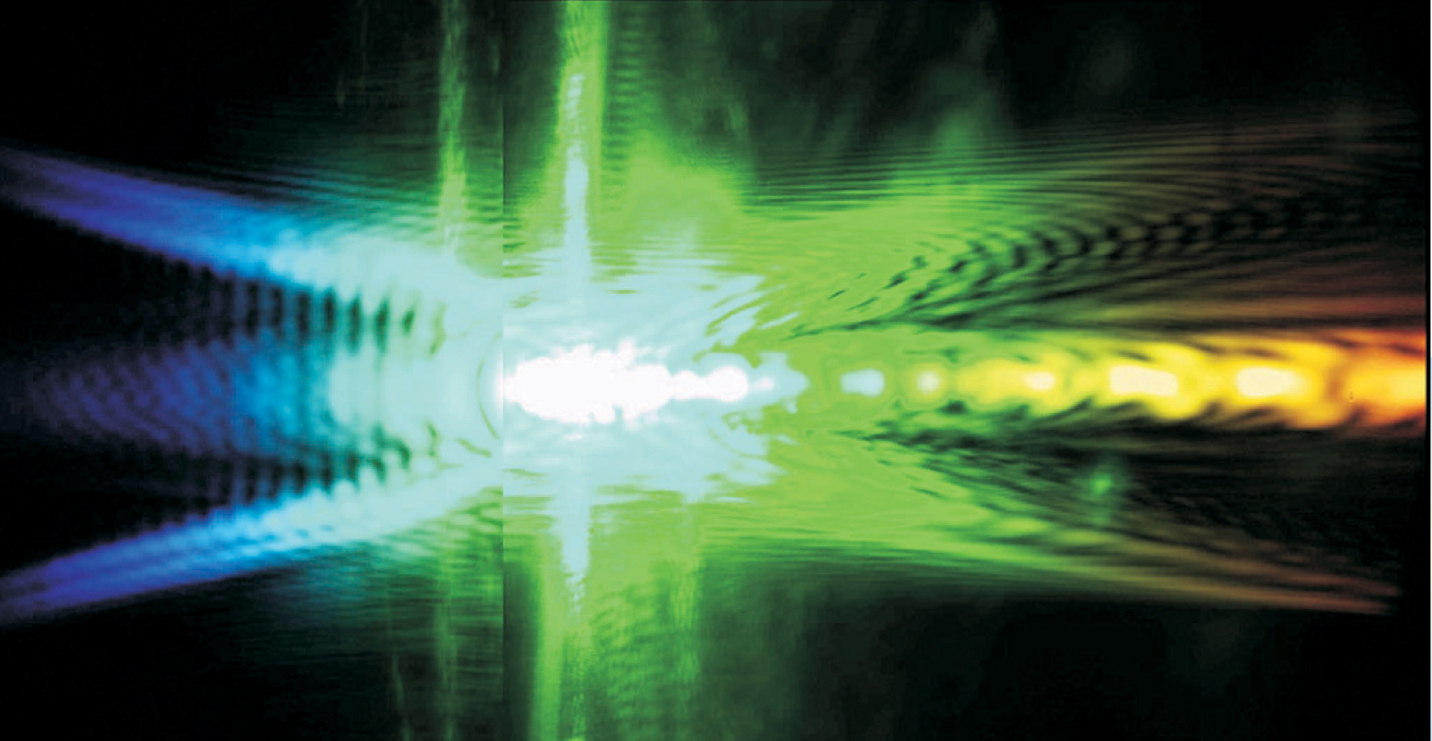
In the Como experiment, the pulses emerged from the laser aperture with the usual Gaussian profile. But when they encountered the dispersive medium of the crystal, nonlinear interactions spontaneously transformed them into a new shape that in longitudinal cross section looks like an X. Figure 1 shows an example.

Nonlinear X waves retain their characteristic profiles when measured in Fourier space. To make this image, an X wave was sent through a lens and onto a spectrograph. The horizontal axis is angular frequency and the vertical axis is wave number.
(Courtesy of Paolo Di Trapani.)
Nonspreading “X waves” had been created before, but in the lower-power linear regime using special equipment. In the nonlinear regime, by contrast, the X wave appears to embody a naturally occurring mode of the light–matter interaction.
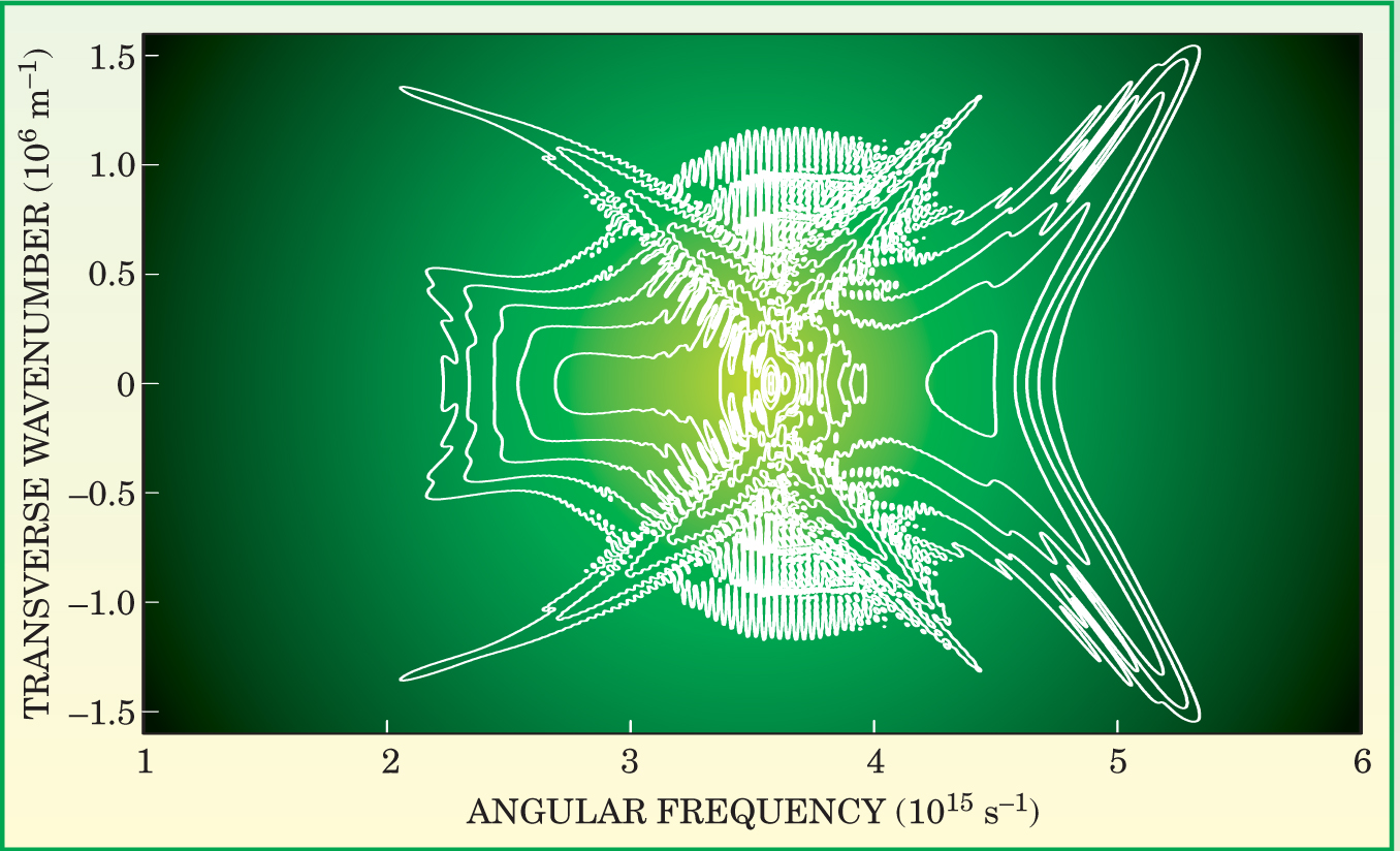
More recent experiments support that emerging view. Di Trapani’s team has found that nonlinear X waves form spontaneously and propagate without spreading in the unexotic medium of water. 2 The observation inspired Miroslav Kolesik, Ewan Wright, and Jerry Moloney of the University of Arizona in Tucson to bring their computer model to bear on X waves. Viewed in silico at a resolution far higher than is available in vivo, the X wave appears not as a stable entity, but as an attractor-like object about which chaotic configurations continually form and reform. 3 Figure 2 shows the attractor in Fourier space.

Computer simulation of an X wave traveling through water. At any instant in real time, the wave would look chaotic. But in the Fourier transformed space, the wave has a distinct, X-shaped profile and resembles the laboratory image shown in figure
(Adapted from ref. 3.)
X waves make up only one of several paths on the quest to create nonspreading beams or pulses of light. Such beams and pulses, the questers hope, could one day underlie new applications in microscopy, fabrication, and communication. The X wave story, however, began with a purely intellectual pursuit: solving an equation.
Bessel beams and ultrasound
In 1987, James Durnin of the University of Rochester in New York discovered a family of nonspreading solutions to the Helmholtz wave equation. The simplest family member consists of a monochromatic beam whose radial profile is the zeroth-order Bessel function.
To make the wave, Durnin, along with his Rochester colleagues Joseph Miceli and Joseph Eberly, built an apparatus consisting of a narrow circular slit and a convex lens. 4 Laser light passing through the slit diffracts, interferes with itself, and, after passing through the lens, transforms into a freely propagating beam.
Viewed from the side, the “Bessel beam” consists of a bright spot from which two hollow cones of light open, forward and backward. As the wave propagates, the central spot is continuously replenished by the constructive interference of the wave’s extended conical components.
In theory, the bright spot requires two infinite conical halos to sustain its propagation, but the Rochester team found that even with an aperture of a few millimeters, the Bessel beam traveled without spreading over a longer distance than did a Gaussian beam.
Among the readers of Durnin’s paper was Jian-yu Lu, a postdoc at the Mayo Clinic in Rochester, Minnesota. In Durnin’s nonspreading Bessel beam, Lu saw the potential for extending the focal depth of ultrasound imaging.
Ultrasound images, like radar maps of approaching aircraft, are formed by sweeping a pulsed beam through the region of interest. Lu, together with Mayo’s James Greenleaf, found a way to create short pulses from the superposition of Bessel beams of different frequencies. 5 Because of their distinctive profiles, Greenleaf named the pulses X waves.
Lu and Greenleaf’s superposition scheme, which was published in 1992, works because the chromatic dispersion of ultrasound in water is negligible. That’s not the case for light in most condensed media. In 1997, Heiki Sõnajalg, Margus Rätsep, and Peeter Saari of the University of Tartu in Estonia figured out how to counter chromatic dispersion and create optical X-wave pulses. 6
The Tartu setup included a holographic element, the “lensacon,” which distributes the frequency components of an X-shaped pulse about different, adjustable cone angles. Using the setup, the Tartu team sent 210-fs pulses through 7 cm of transparent dielectric. The pulses crossed the sample without spreading.
Going nonlinear
Initially, Di Trapani didn’t set out to make nonlinear X waves. Rather, he, like many others, was working with the prevailing paradigm for localized optical waves in the nonlinear regime: the soliton. Solitons propagate without spreading, thanks to a nonlinear phase shift that compensates for dispersion.
Optical solitons have been created in one-dimensional systems, such as optical fibers, but localizing them in three dimensions requires higher intensities, which, through nonlinear losses, tend to quench the soliton.
In 1998, Di Trapani teamed up with Algis Piskarskas’s group at the University of Vilnius in Lithuania. Their aim was to come up with a way of making sure a second harmonic would travel with its fundamental in a soliton-like package despite the dispersion-induced difference in group velocity. Their solution was to “tilt” the pulses—that is, to bounce them at an angle off a grating to give the faster colors a larger cone angle and, consequently, a lower effective speed. 7
The tilting trick worked with plane waves and yielded temporal solitons—that is, waves localized in the propagation direction. But in the transverse plane, the waves are exquisitely sensitive to fluctuations—even to quantum noise. The instability arises because of the intensity dependence of refractive index. Bright light travels more slowly than dim light. As a beam propagates, its dim outer part will tend to lead—and, therefore, collapse around—the slower hot spot, an effect known as Kerr focusing.
Achieving 3D localization requires figuring out how to counteract spreading while preventing collapse. When the chromatic dispersion is anomalous—that is, when refractive index decreases with frequency—symmetric Kerr focusing occurs in the spatial and temporal domains and leads to strong nonlinear dissipation and collapse. But when chromatic dispersion is normal, as is the case for optical light in most transparent media, broadening occurs in the time domain and collapse is forestalled.
The tilted pulse experiments showed that angular dispersion can compensate for the broadening in the nonlinear regime. Di Trapani realized that X waves, the radial generalization of tilted pulses, might avoid both time broadening and collapse. In 1999, he and Piskarskas set themselves the goal of making nonlinear X waves and formed a collaboration with colleagues at the universities of Ferrara, Rome, and Madrid.
In the linear regime, the Tartu group had used its lensacon to impose the requisite angular dispersion. Working in the nonlinear regime, Di Trapani and his collaborators saw hints that different colors seemed to find the right, dispersion-defeating angle by themselves.
Those preliminary observations in 2001 led the team to predict the existence of localized X waves in the nonlinear regime. Computer simulations and the implementation of a three-dimensional mapping diagnostic confirmed the prediction.
Nonlinear X waves, it appears, arise from a spatiotemporal instability that is intrinsic to the medium. The instability shows up dramatically in the high-resolution Tucson computer simulations. By switching on and off various physical phenomena in their model, Kolesik, Wright, and Moloney confirmed Di Trapani’s finding: The interplay of Kerr nonlinearity and chromatic dispersion is responsible for the nonspreading propagation of X waves.
Missed tails
Physicists have been firing lasers at crystals for decades. Why did no one notice the spontaneously forming X waves? Di Trapani, who missed the waves himself, suggests several reasons.
Nonlinear X waves are intrinsically spatiotemporal phenomena. A theoretical approach that separates space and time will fail to capture X waves. It’s also a mistake, Di Trapani believes, to regard the linear and nonlinear regimes as separate.
An X wave is more centrally concentrated than a Gaussian beam, but the two would look similar in a detection scheme designed to track a beam’s full width at half maximum. Only the weak extended halos of the X wave, easily dismissed as noise, might betray the waves’ presence.
The weak halos of an X wave sustain its propagation but, in the linear case, have so far limited X-wave applications. Lu and Greenleaf found that the halos acted as a source of blur that vitiated the bright spot’s deep focusing.
In nonlinear X waves, the halos are much dimmer than the bright spot. Indeed, the halos lie in the linear regime. Recently, Di Trapani has found that an imbalance develops between the leading and lagging halo. The imbalance helps steel the X wave against nonlinear losses that would otherwise quench it.
Whether the favorable properties of nonlinear X waves will lead to applications is not clear. However, as a paradigm for describing the nonlinear interaction of light in dispersive media, they are already proving useful.
References
1. P. Di Trapani et al., Phys. Rev. Lett. 91, 093904 (2003).https://doi.org/10.1103/PhysRevLett.91.093904
2. A. Dubietis, E. Gaizauskas, G. Tamošauskas, P. Di Trapani, Phys. Rev. Lett. 92, 253903 (2004).https://doi.org/10.1103/PhysRevLett.92.253903
3. M. Kolesik, E. M. Wright, J. V. Moloney, Phys. Rev. Lett. 92, 253901 (2004).https://doi.org/10.1103/PhysRevLett.92.253901
4. J. Durnin, J. J. Miceli, J. H. Eberly, Phys. Rev. Lett. 58, 1499 (1987).https://doi.org/10.1103/PhysRevLett.58.1499
5. J. Lu, J. F. Greenleaf, IEEE Trans. Ultrason. Ferroelectr. Freq. Control 39, 19 (1992).https://doi.org/10.1109/58.166806
6. H. Sõnajalg, M. Rätsep, P. Saari, Opt. Lett. 22, 310 (1997).
7. P. Di Trapani et al., Phys. Rev. Lett. 92, 253093 (1998).


