Attosecond Bursts Trace the Electric Field of Optical Laser Pulses
DOI: 10.1063/1.1825259
Last year, Ferenc Krausz, Theodor Hänsen, and their collaborators used femtosecond laser pulses to make isolated attosecond bursts of UV light. 1 To pull off the feat, the team wielded precise and reproducible control over the five or so electric field oscillations that fit inside the amplitude envelope of a femtosecond pulse. With the right phasing between field and envelope, one specific oscillation in each pulse spawned an attosecond burst on demand (see Physics Today, April 2003, page 27 ).
Getting the phasing right depended on measuring the bursts’ UV spectrum, which was fed back into a sophisticated setup of amplifiers, interferometers, and other equipment. But now Krausz, who has joined Hänsch at the Max Planck Institute for Quantum Optics in Garching, Germany, can see the phasing directly. In a new experiment, he and a group led by Ulrich Heinzmann of the University of Bielefeld in Germany have used attosecond bursts to map the electric field of femtosecond pulses—the same pulses that begat them. 2
Electromagnetic waves have been mapped before, in the terahertz regime, using a technique called electro-optical sampling (EOS). Developed in the 1980s, EOS relies on the Pockels effect, the dependence of the polarization of certain crystals on an applied electric field. As a terahertz wave travels through a Pockels cell, the change in polarization induced by the wave’s electric field can be probed with an optical pulse. Varying the time delay between the terahertz wave and the optical pulse maps the terahertz electric field as a function of time.
EOS can’t resolve optical waves. Because the probe pulses must be shorter than the wavelengths under investigation, mapping an optical wave would require an extreme UV probe. In that waveband the transparent crystals in Pockels cells are uselessly opaque.
Krausz’s mapping method eschews light as a probe. Instead, it uses an attosecond burst to create a precisely timed flash of photoionization in a gas just as a femtosecond pulse passes through. The electric field of the pulse accelerates the photoelectrons, which, depending on the pulse phase, are either hastened or delayed on their journey to a time-of-flight detector. The TOF measurement captures information about the field at the instant of ionization; repeating the measurement on identical pulses at different time delays captures the whole field.
A new challenge
Krausz and his collaborators can routinely create identical femtosecond pulses and precisely timed attosecond bursts, although the procedure remains complex and difficult. To measure the electric field of the femtosecond pulses, the team faced a new challenge: to ensure the photoionization occurs at an adjustable value of the field’s phase. Here’s how they do it.
First, they shoot femtosecond laser pulses into a target of neon atoms to produce bursts of 93-eV photons that last 250 attoseconds. The pulses and bursts travel together in a collimated beam, but the pulses, because of their longer wavelength, diverge more than the bursts. That difference is crucial and comes into play when the bursts and pulses reach a specially prepared two-part mirror. Whereas the pulses spread over a 25-mm-wide circular region of the mirror, the bursts are confined to a much smaller concentric spot. By moving the small central part of the mirror back and forth, the experimenters can adjust the delay between the bursts and the pulses. The nanometer precision of the mirror’s translation stage corresponds to attosecond precision in time delay.
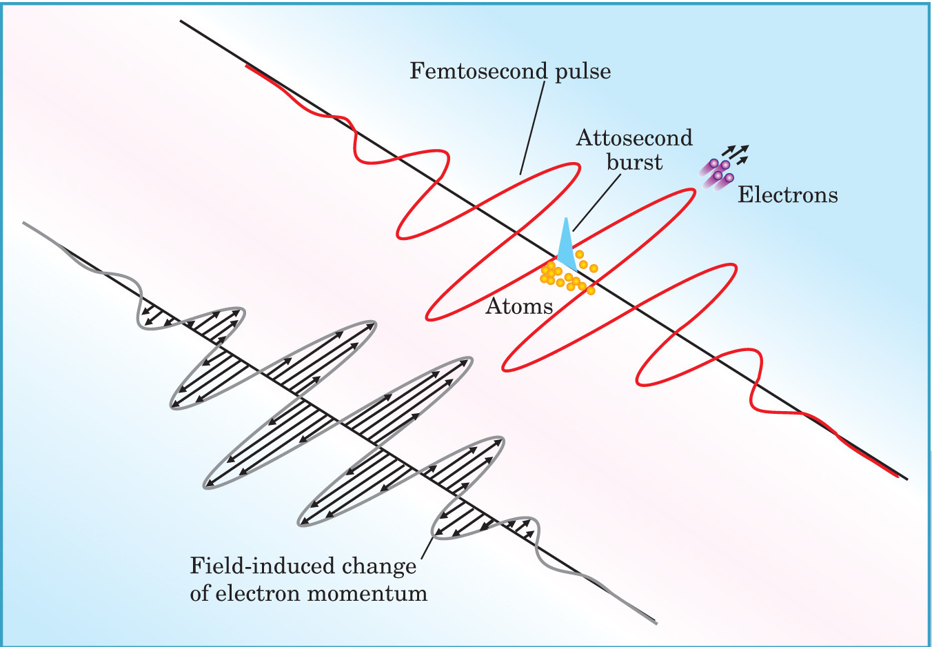
Figure 1 shows schematically what happens next. After bouncing off the mirror, the pulses and bursts converge on a second neon target. The pulse photons lack the intensity to ionize the gas, but the burst photons each pack enough energy to ionize a neon atom. Once freed, a photoelectron acquires a momentum kick per unit charge that equals the time integral, from the time of release to infinity, of the electric field of the pulse—that is, the vector potential.

When an attosecond burst (blue) ionizes neon atoms, photoelectrons receive a momentum kick (black arrows) determined by the electric field (red) at the time of the burst.
(Adapted from ref. 2.)
On average, the TOF detector captures only about 1 photoelectron per pulse. But the high firing rate of the laser and robust reproducibility of the pulses and bursts ensure that a complete picture of the pulse field can be put together.
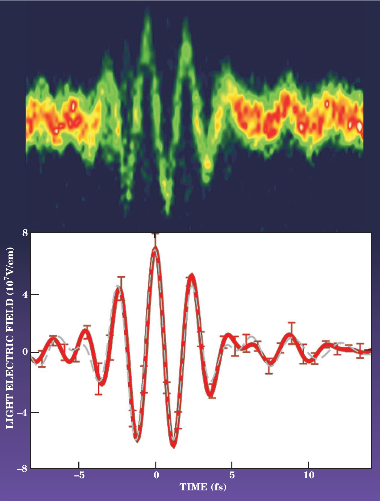
That’s what appears in figure 2. The top panel shows kinetic energy spectra of the detected electrons. Because the kinetic energy the photoelectrons acquire from the electric field is small compared to their initial energy, the change in kinetic energy is proportional to the momentum kick—and hence to the field’s vector potential. The lower panel shows the field itself, obtained by taking the time derivative of the vector potential. As the units on the y axis attest, the experiment yields not only the variation of the electric field, but also its absolute strength.

Time-of-flight data trace the electric field of the femtosecond pulses. The upper panel shows the kinetic energy spectra in 200-attosecond-wide vertical slices as a function of time delay. The dynamic range of the spectra is large enough and time step small enough that numerically differentiating the vector potential yields the field, which is shown in the lower panel.
(Courtesy of Eleftherios Goulielmakis, Max Planck Institute for Quantum Optics.)
Doubtless the shape of the field traced in figure 2 would not surprise James Clerk Maxwell or Heinrich Hertz—let alone a 21st-century physicist. But the figure embodies an unprecedented degree of control over light on the attosecond timescale. Columbia University’s Tony Heinz points out that major developments in such fields as communications and magnetic resonance imaging resulted from the precise control of electric fields in the RF regime. He adds, “This new step of full amplitude and phase control of electromagnetic radiation at much higher frequencies offers great potential for both scientific and technological advances.”
References
1. A. Baltuška et al., Nature 421, 611 (2003).
2. E. Goulielmakis et al., Science 305, 1267 (2004).https://doi.org/10.1126/science.1100866


