Detecting Illicit Radioactive Sources
DOI: 10.1063/1.1839375
Concerns about the illicit movement of radionuclides across national borders have heightened the degree of protection that the US and western European countries have instituted. The challenge is not only to detect hidden radioactive materials, but also to distinguish them from legitimate radionuclides such as radiopharmaceuticals that are often transported across borders and shipped throughout a country. Every day, more than 300 000 vehicles, roughly 2500 aircraft, and nearly 600 ships pass through US ports of entry. With more than 600 US border sites to protect, screening imported radioactive material requires a careful balance of high throughput and high search efficiency. Unfortunately, the two requirements are at odds: Rapid screening implies less counting time available to detectors (see the article by Jay Davis and Don Prosnitz in Physics Today, April 2003, page 39 ).
What complicates the efficiency of this screening process is that many common imported goods are either intentionally or naturally radioactive. Commercial shipments of ceramic-glazed materials, abrasives, road salt, and even kitty litter, for example, contain naturally occurring radionuclides that may trigger false alarms from radiation detectors. Moreover, medical radionuclides are used throughout the US, so the problem of detecting potentially illicit radioactive sources is not confined to border crossings. Approximately 120 000 people in the US can be expected to carry a detectable radioactive trace due to diagnostic and therapeutic procedures they’ve undergone.
In 2003, the International Atomic Energy Agency (IAEA) convened a conference 1 on the potential problems presented by radiological dispersal devices, or dirty bombs. Secretary of Energy Spencer Abraham argued for an improvement in US technology designed to quickly detect radioactive materials that would pose a threat in a terrorist’s hands. And other attendees identified several radionuclides of particular concern: cobalt-60, strontium-90, cesium-137, iridium-192, plutonium-238 and -239, and americium-241. To uncover the presence of such radionuclides, instruments must be capable of detecting emitted radiations at a distance of meters from the source and probably after the radiations have passed through intervening material. Several of those isotopes emit penetrating gamma rays with energies in the range of 50 to 1300 keV, while others, such as 90Sr, emit beta particles that have relatively short ranges, a problem that makes them more difficult to detect. Bremsstrahlung photons produced when the 90Sr beta particles interact with high-atomic-number materials will have a higher probability of detection due to their longer ranges.
The US, Russia, and other nations store large quantities of special nuclear materials (SNMs)—fissionable radionuclides such as 239Pu and the 233 and 235 isotopes of uranium—in protected locations, although the threat exists that such material could be stolen and fashioned into a nuclear device. Plutonium isotopes emit alpha particles, gamma rays, and characteristic x rays released in atomic transitions of bound electrons. Some of these x rays have energies below 30 keV, which makes them difficult to detect from a distance or if shielded. Neutrons are also emitted by these radionuclides as a result of spontaneous fission, and α,n interactions with surrounding materials.
Like the low-energy x rays, alpha and beta particles emitted by the actinides and other high-risk nuclides have little penetrating power; their energies are just a few MeV and their detection probability at the exterior of any container is nearly negligible. The radiations most amenable to detection at reasonable distances are gamma rays because of their exponential absorption in matter. The photoelectric and Compton interactions are dominant in the interactions of matter with photons that have energy below about 1 MeV. Secondary electrons produced from the collisions of gamma rays with atomic electrons can deposit their energy in materials such as gases, crystals, plastics, or organic liquids and may produce an avalanche of free electrons, electron–hole pairs, or light pulses, depending on the detector material and construction.
New tools
In the laboratory, large, cryogenically cooled devices can sensitively identify the isotopic signatures of various emissions. However, those instruments were never intended to deal with the problem of searching for SNM in the large volume of traffic encountered on public highways, airports, and seaports. The threat of terrorism has prompted researchers and manufacturers to develop new types of portable and more flexible instruments.
Four basic types of such portable instruments now exist. Personal radiation detectors (PRDs) are small, highly sensitive devices, about the size of a cell phone, that generally incorporate an inorganic scintillator (converting high-energy photons to visible light) coupled to a photomultiplier tube along with electronic circuitry to count pulses and perhaps correct for background radiation; some can even detect both photons and neutrons. For emergency first responders, for instance, PRDs provide a reliable alert to radiation levels at some preset threshold.
Hand-held survey meters similar to those found in nuclear power plants to measure exposure or dose equivalent rates can also be used to search for radioactive material. With slightly more sophisticated electronics than PRDs—better read-out capabilities, for instance—these meters may also contain interchangeable probes consisting of scintillation detectors and phototubes, and large-volume (or high-pressure) ionization chambers or neutron detectors.
Similar in size to survey meters, radionuclide identifier devices (RIDs) often use a sodium iodide crystal, photomultiplier, and associated electronics to produce a pulse-height spectrum. In some instruments, the data can be compared to a stored library of spectra to identify the radionuclides. The difficult task of resolving the nuclides is normally performed using a cooled high-resolution detector, pulse-processing electronics, a multichannel analyzer, and a computer. The need to miniaturize a lab system, use batteries, and keep size, weight, and cost low inevitably compromises performance. Nevertheless, the hand-held meters provide valuable information to help discriminate between false alarms from naturally occurring or medical isotopes and real ones from high-risk radionuclides. A commercial instrument that was recently introduced uses a Stirling-engine cooler and portable high-purity germanium detector; the continued development of such devices will undoubtedly result in newer, smaller, less-expensive models.
The fourth and largest detector type is the radiation portal monitor (RPM). Large, usually plastic scintillators such as polyvinyl toluene make up these detectors, which are coupled to phototubes, mounted in moisture-resistant enclosures, and placed on either side of a roadway, as shown in figure 1. These devices primarily detect photons. Some plastic scintillators can also detect neutrons, but more often, RPMs with large 3He gas-filled detectors are used for that job.

Figure 1. Highly sensitive portal monitors (yellow) installed in passenger lanes at the Pacific Highway crossing in Blaine, Washington, can detect the presence of small amounts of radioactive material just meters away. False alarms occur somewhat frequently—most often from naturally radioactive agricultural products, ceramic tiles, and the rare passenger who may have just received a thallium, technetium, or iodine injection from a physician.

Key contributors
Historically, a variety of research communities have advanced the field of radiation detection for their distinct purposes. In the particle physics community, the need to measure a wide range of subatomic particles was met by pioneers such as Georges Charpak, who received the 1992 Nobel Prize in Physics for his invention and development of particle detectors—in particular, the multiwire proportional chamber, introduced in 1968. Since then, particle physicists have developed scintillation detectors to study a wide range of radiations produced at accelerators worldwide.
At the low-energy end of the spectrum, diagnostic nuclear medicine developed segmented detectors to provide positional, imaging information from reconstruction of the photon transmission paths in the human body. Recently, medical engineers combined this computed tomography with positron emission tomography in a single unit. Both CT and PET detectors are optimized to record photons from 80 keV up to 511 keV, the energy of annihilation radiation. Because of the high effective atomic number needed to detect the 511 keV photons efficiently, bismuth germinate and other inorganic scintillators are now widely used.
To monitor the effluents of nuclear power plants for radionuclides that may be released to the environment, radiochemists use gamma-ray spectrometers and analysis software to deconvolve the fission and activation products from complex spectra with lines in the 20 keV to 2 MeV range. This kind of work has driven the development of large and high-resolution hyperpure germanium gamma-ray spectrometers. And the software that sorts through spectra to list component radionuclides is now commercially available.
Weapons inspectors and others in the safeguards community use photon spectrometry to detect and identify nuclear materials, but with a focus on the low-energy x rays characteristic of the actinides and a few associated gamma-ray lines. This community developed much of the technology now found in PRDs, RIDs, and RPMs to monitor specific locations that contain SNMs; they also incorporated software that analyzes emissions in the energy range typical of uranium and plutonium isotopes.
Weapons inspectors rely on instruments and techniques similar to those used for low-level radiochemistry measurements at nuclear power plants. The mixed-nuclide spectra may come from gaseous samples (mixed radioxenons and kryptons) or soil samples taken near nuclear facilities. The main requirements for the detectors used by laboratories are high photon-detection efficiency and high-energy resolution.
Measuring the neutron fluence and the neutron energy spectrum has long been a concern of health physicists because a detailed knowledge of both is required to accurately calculate the radiation effects of neutrons on biological tissue. Radiation protection dosimetry measurements for astronauts and workers at high-energy accelerators and nuclear power plants require a combination of detectors to sort out the risks from fast and thermal neutrons and from photons and charged particles. Personnel dosimeters are used to determine the dose equivalent for radiation workers. These often use thermoluminescent materials to detect photons, and etched-track devices made of plastics to detect neutrons. Passive dosimeters require subsequent processing after exposure and are not useful for the immediate detection of radiation.
Researchers conducting solar neutrino physics experiments consider fast neutrons to be a particular nuisance. Distinguishing those neutrons from more interesting data has led to very sensitive methods of measuring low neutron fluences in the presence of high gamma-ray backgrounds. Schemes for neutron–gamma discrimination rely on pulse-shape discrimination or on coincidence signals from multiple detectors. The hardware and software advances in handling data from neutron scintillators may be readily incorporated into the next generation of sensitive neutron counters for nuclear materials.
There are two areas of concern when dealing with illicit radioactive sources: detection and protection. Different quantities and units are used in each field. Radiochemists and safeguards workers are comfortable with quantities such as activity and radionuclide mass, whereas those in health physics deal with the dose equivalent and dose equivalent rate. For a primer on how the quantities and units of one field can relate to another, see the
Gamma-ray detectors
Among the earliest scintillators used to detect ionizing radiation were organic compounds such as naphthalene (C10H8) and anthracene (C14H10), in combination with photomultipliers that amplified their light output. 2 But today, sodium iodide activated with 1% thallium is the most frequently employed compound used to measure gamma rays, 3 partly because of its high-atomic-number iodine content, which increases photon detection efficiency. Other high-atomic-number materials used successfully include thallium-doped cesium iodide, bismuth germanate, and cadmium zinc telluride; recently, cerium-doped lanthanum chloride and lanthanum bromide 4–6 have been investigated. Lithium iodide can also be used to detect gamma rays, but it is often used to detect thermalized neutrons because of lithium’s high neutron cross section.
These inorganic scintillators produce light pulses as a result of interaction of gamma rays with atoms in the lattice. The high-energy photons kick out core- or valence-shell electrons, which subsequently give off lower-energy visible light when they recombine with holes left behind. When added in low concentration to the melt used to grow the crystal, elements such as thallium or europium create lattice vacancies that alter the electronic structure and control the photon wavelengths emitted—and thus create one of the properties needed for an effective scintillator.
Good scintillators produce large light pulses, whose intensities are proportional to the incoming photon energy. They must also be transparent to the wavelengths produced so that the light can escape, be detected, and be amplified in a later photomultiplier stage.
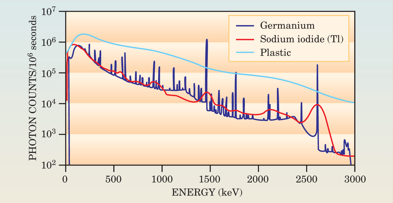
Figure 2 illustrates the difficulty of identifying a radionuclide using the various types of scintillators commonly found in the instruments that now search for high-risk radionuclides. In the laboratory, pure germanium provides a spectrum that clearly shows the photon energy peaks. But materials used in the field—NaI(Tl), for instance, and the large plastic scintillators commonly used to monitor borders—have poorer resolution, which makes it more difficult to discern the identity of any radionuclides detected.

Figure 2. These spectra indicate the relative sensitivity of three different materials to the natural background gamma rays present in southeastern Washington. The high-resolution germanium detector (blue) outperforms the thallium-doped sodium iodide detector (red), whose resolution is too limited to reliably identify as many individual radionuclide spectral lines. Large plastic scintillators (light blue) of the type used in a portal monitor system are an order of magnitude more sensitive, but even more unsuitable for energy discrimination.
(Data courtesy of W. K. Hensley, Pacific Northwest National Laboratory, Richland, WA.)
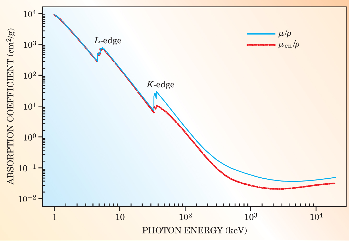
Moreover, many of the photons emitted by the materials of concern to emergency responders occur in an energy region where the scintillator’s absorption coefficients are changing rapidly. So their response as a function of incident photon energy will also vary significantly. The plot in figure 3 shows the mass attenuation and mass-energy absorption coefficients for CsI as a function of energy. 7 The large changes occur in the 1–500 keV range where the photoelectric absorption coefficient decreases, while the Compton interaction coefficient begins to increase and becomes the dominant contribution above about 500 keV.

Figure 3. The detection capability of inorganic scintillators such as thallium-doped cesium iodide depends on their radiation absorption properties. This plot shows the mass attenuation coefficient µ/ρ and mass-energy absorption coefficients µen/ρ as a function of photon energy. The coefficient µ/ρ is akin to the exponential absorption coefficient and µen/ρ reflects the photon energy transferred to secondary electrons. Because the absorption coefficients rise rapidly due to the predominance of photoelectric interactions as photon energy decreases, the response of Csl(Tl) scintillators necessarily increases for lower-energy photons.
(Data courtesy of J. H. Hubbell and S. M. Seltzer, NIST; available at http://physics.nist.gov/PhysRefData/Xray-MassCoef/ComTab/cesium.html.)
Due to the attenuation in any surrounding shielding, however, the fluence as a function of incident gamma-ray energy for these detectors will generally peak at approximately 60 keV and then fall off at lower energies. Therefore, the sensitivity of the scintillator-based instruments is relatively large for photons such as those emitted by 241Am and plutonium isotopes.
Neutron detectors
Californium-252 is occasionally used as a source of neutrons for in-patient cancer radiation therapy, and some industrial moisture gauges employ 241Am–Be sources. Cosmic-ray-produced neutrons are also present as weak background radiation, but otherwise, neutron sources are scarce in everyday activities. So, if a neutron detector triggers an alarm at an airport or border crossing, it represents a situation of some concern because it could indicate the presence of a nuclear weapon.
To reliably register the presence of enriched uranium or plutonium, an instrument must detect a small fluence of fast neutrons and provide an energy spectrum fine enough to discriminate between a 241Am–Be source used to confirm the presence of oil or water in an exploratory well, say, and weapons-grade plutonium. Although routine in the laboratory, these measurements are a tall order for a battery-powered RID. At the moment, no portable commercial instrument exists that can make reliable identifications of neutron spectra within seconds.
Because neutrons are indirectly ionizing particles, they interact by elastic and inelastic nuclear scattering to produce secondary charged particles. Gaseous and solid state detectors or organic scintillators can detect those charged particles. 6LiI or 3He gas both have neutron-reaction cross sections that vary inversely with energy. For detection purposes, the moderation of fast neutrons using a material such as polyethylene—which produces lower-energy neutrons from elastic scattering—makes it possible to take advantage of the large thermal-neutron absorption cross sections. Consider the following reactions:
6Li(n,α)3H ( Q = 4.78 MeV; σth = 940 barns);
10B(n,α)7Li ( Q = 2.31 MeV [93%] or 2.79 MeV [7%]; σth = 3837 barns;
3He(n,p)3H ( Q = 0.764 MeV; σth = 5333 barns).
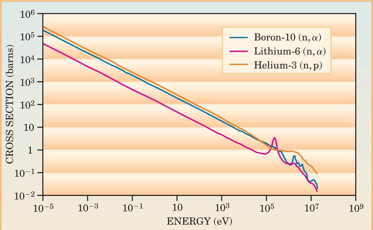
Here, Q is the energy released in the reaction 8 and σth is the thermal-neutron absorption cross section. 9 In this notation, a neutron incident on a 6Li atom releases an alpha particle to produce 3He, for example. The high sensitivity of lithium, boron, and helium to slow neutrons is advantageous considering the low neutron-fluence rates routinely encountered. On the other hand, the decrease in magnitude of the cross sections at higher energies makes the detectors relatively insensitive to fast neutrons (see figure 4). Personal radiation detectors that are held close to the body may respond to moderated and reflected neutrons having a lowered energy and hence a greater likelihood of being sensed by a an inverse energy detector.

Figure 4. To detect low-energy neutrons one usually employs one of three reaction targets—3He, 10B, and 6Li—whose cross sections are shown. The very large cross sections (1 barn = 10−28 m2) below about 10 eV provide the detector’s sensitivity. But designers have to moderate higher-energy neutrons to take advantage of these large cross sections. In that process, the practicality of the detector is somewhat compromised since the amount of moderating material necessary makes the detectors bulky and heavy.
(Neutron cross section data from the National Nuclear Data Center, Brookhaven National Laboratory, www.nndc.bnl.gov.)
Neutrons are nearly always accompanied by photons, and in many cases the photon contribution to the fluence spectrum will be dominant. So instrument designers have to pay close attention to the sensitivity of neutron detectors to photons of various energies. Because some radiation detection instruments are worn on the belt, neutrons entering the body can cause specific reactions that generate photons—in particular, the neutron capture reaction in hydrogen, 1H(n,γ)2H, which produces a 2.2-MeV photon.
Gaseous detectors that produce a pulse-height spectrum can sometimes discriminate between neutrons and photons. It may be possible to discriminate against photons simply by setting an electronic threshold above which photon-induced events are unlikely, for instance. Moderator-based neutron area-survey instruments often use that technique. Applying pulse-shape analysis may also be possible, if the pulse shapes are distinguishable, as they are, for example, in organic-crystal or -liquid scintillators.
A relatively recent development is the superheated-emulsion detector, which uses small droplets of superheated liquid suspended in a viscoelastic medium to detect neutrons. A fast neutron that interacts with a nucleus near one of those droplets can cause localized evaporation and the formation of a small vapor bubble. Optical or acoustical methods can then detect the bubbles and in some cases also provide enough detail to deduce some spectrometric information. 10
In general, large-volume neutron detectors are more effective than emulsion detectors, but the need for a device small enough for a pocket or belt makes it difficult to incorporate a large neutron detector. The development of more efficient small devices is therefore a subject of significant interest, and a number of programs are under way at national laboratories, universities, and industrial research facilities. Currently available portable commercial instruments have limited neutron-detection capabilities.
Homeland security
The establishment of the Department of Homeland Security (DHS) signaled the US government’s commitment to mitigate terrorist threats. Emergency workers, such as firefighters, police, HAZMAT teams, customs officials, border-protection personnel, and Coast Guard staff, are being issued radiation detectors and radionuclide identifiers and are being trained in their use at airports, seaports, and land borders.
Even before it was formally created, staff members of what became DHS requested standards to evaluate the new instruments needed to detect and interdict illicit radioactive materials. The American National Standards Institute (ANSI) developed the new standards, which cover personal radiation detectors, hand-held survey meters, portable radionuclide identifiers, and portal monitors, in record time with representatives from national laboratories, instrument manufacturers, and university researchers. 11 The standards will form the basis of performance tests carried out in the next few months and be used in a database for emergency response organizations that decide on new equipment purchases. That equipment purchased with DHS funding will itself have to undergo ANSI standards-based testing; the goals are to confirm instrument performance and drive product improvement. DHS is also working to coordinate standards development and instrument performance testing with other governments. 12,13
A brief summary of the tests required in the new standards includes
resistance to mechanical shock and vibration
battery lifetimes
effects of environmental influences such as electromagnetic and radio frequency interference
effects of temperature and humidity
detection sensitivity and speed of response.
These type tests 11 are intended for specific models of commercial instruments and are meant to simulate the broad range of environmental conditions encountered in actual field use. The goal, of course, is to check that instruments used by emergency responders perform reliably.
Quantities and Units
Because physicists in different fields have separately contributed to detector technology, different quantities are often used for radiation detection and radiation protection. Emergency responders require an unambiguous measure of what levels of radiation constitute a hazard following an accident or terrorist attack. The problem is, radioactive material is often measured in terms of the activity of a source, or its mass, whereas the quantities that indicate the degree of radioactive exposure a human being may have suffered are given as the dose equivalent (see article by Bert Coursey and Ravinder Nath, Physics Today, April 2000, page 25 ).
The dose equivalent H is the product of the quality factor Q—a measure of the biological effect of the radiation—and the absorbed dose D at a certain point in the tissue. Q(L), for instance, expresses the quality factor at a point after a particle’s initial energy has been attenuated through a distance L in the body. The quality factor for photons is 1; the quality factor for neutrons varies from approximately 3 to 20, with the maximum value occurring at about 0.5 MeV. The unit for dose equivalent is known as the sievert (Sv), given by units of J kg−1.
The other dosimetric quantity, air kerma (K), can be related to the activity of a photon-emitting source. Air kerma is defined as dE tr/ dm, where dE tr is the sum of the initial kinetic energies of all the charged ionizing particles liberated by uncharged ionizing particles within a mass dm of dry air. The unit for air kerma is known as the gray (Gy), given by units of J kg−1.
To relate the different dosimetric quantities, consider the air-kerma rate constant Γδ, given by
The air-kerma rate, essentially proportional to the quantity registered by a simple exposure-rate meter for a given activity of radionuclide, can be computed for specified radionuclide sources by using the air-kerma rate constants or coefficients given in reference . The relation between air kerma and dose equivalent is in general complex, depending on incident photon energy and angle and upon the organ or the depth within the body, but operationally, the dose equivalent (in Sv) for photons with energies from about 20 keV up to tens of MeV is roughly (within a factor of 2) numerically equal to the air kerma (in Gy).
Research expectations
The Homeland Security Advanced Research Projects Agency (HSARPA) is encouraging researchers at universities and industrial facilities that partner with government labs to develop detectors for radiological and nuclear countermeasure systems. Some of the technical projects the agency expects to see developed in that effort include enhanced radiographic imaging systems; methods to improve discriminating between naturally occurring radioactive materials and those that pose a threat, such as SNMs; improved techniques to identify radionuclides; mobile detection systems; and communication and archiving of detector data.
Radiological imaging systems are expected to use both active and passive techniques to survey a wide variety of vehicles and containers. Los Alamos National Laboratory has recently developed a novel radiographic imaging technique that uses cosmic-ray-produced muons 14 (see Physics Today, May 2003, page 19 ). The radiation source in that case would always be freely available. Gamma-ray detectors are expected to have improved radiation detection and identification properties as discussed above, and their usability in a variety of field conditions is being enhanced. High-sensitivity neutron detectors with spectroscopic capabilities and gamma-ray discrimination remain on the HSARPA wish list. But the agency expects that improved hand-held radionuclide identifiers with increased sensitivity and advanced spectrum analysis algorithms are on the way. Portal monitors with spectral analysis capabilities are also under development.
Beyond the technical requirements, described in detail on the HSARPA webpage (see www.dhs.gov ), an essential practical requirement remains: The new instruments must become commercially available at a reasonable cost, so that large numbers of them can be purchased and deployed in a short time. By combining practical considerations with technical advances, the work needed to mitigate security threats should proceed at a brisk pace.
References
1. Proc. International Conference on Security of Radioactive Sources, Vienna, Austria, 10–13 March 2003; available at http://www-pub.iaea.org/MTCD/publications/PDF/pub1165_web.pdf .
2. H. Kallmann, R. Warminsky, Ann. Physik 4, 57 (1948).
3. G. F. Knoll, Radiation Detection and Measurement, 3rd ed., J. Wiley & Sons, New York (2000).
4. E. V. D. van Loef, P. Dorenbos, C. W. E. van Eijk, K. W. Kramer, H. U. Gudel, Appl. Phys. Lett. 79, 1573 (2001).
5. E. V. D. van Loef, P. Dorenbos, C. W. E. van Eijk, J. Phys. Cond. Mat. 15, 1367 (2003).
6. P. Dorenbos, J. T. M. de Haas, C. W. E. van Eijk, IEEE Trans. Nucl. Sci. 51, 1289 (2004).
7. J. H. Hubbell, EXCOM Tables , NIST, Gaithersburg, MD (2003).
8. G. Audi, A. H. Wapstra, Nucl. Phys. A 595, 409 (1995).
9. S. F. Mughabghab, M. Divadeenam, N. E. Holden, Nuclear Cross Sections Vol. 1: Neutron Resonance Parameters and Thermal Cross Sections, Part A, Z = 1–60, Academic Press, New York (1981).
10. R. E. Apfel, F. D’Errico, Nucl. Inst. Meth. A 476, 298 (2002).
11. American National Standards Institute, ANSI Standard N42.32–35, ANSI, New York (2003).
12. P. Beck,Illicit Trafficking Radiation Detection Assessment Program, ARCS Report, OEFZ–GS–0002, Austrian Research Centres Seibersdorf, February (2000); see http://www.arcs.ac.at/G/GS/system/itrap .
13. Coordinated Research Project, “Improvement of Technical Measures to Detect and Respond to Illicit Trafficking of Nuclear and Other Radioactive Materials,” Consultants’ Meeting, Vienna, Austria, 17–21 March 2003.
14. K. N. Borozdin et al., Nature 422, 277 (2003).
15. S. M. Seltzer, Air-Kerma-Rate Coefficients for Selected Photon-Emitting Radionuclide Sources, NIST, rep. no. NISTIR 7092, Washington, DC (2004).
More about the authors
Joe McDonald is a laboratory fellow at the Pacific Northwest National Laboratory in Richland, Washington. Bert Coursey is director of standards and Michael Carter is science adviser to the Assistant Secretary for Science and Technology at the Department of Homeland Security.
Joseph C. McDonald, Pacific Northwest National Laboratory, Richland, Washington, US.
Bert M. Coursey, Department of Homeland Security, US.
Michael Carter, Department of Homeland Security, US.


