X-ray ghost imaging
DOI: 10.1063/PT.3.3319
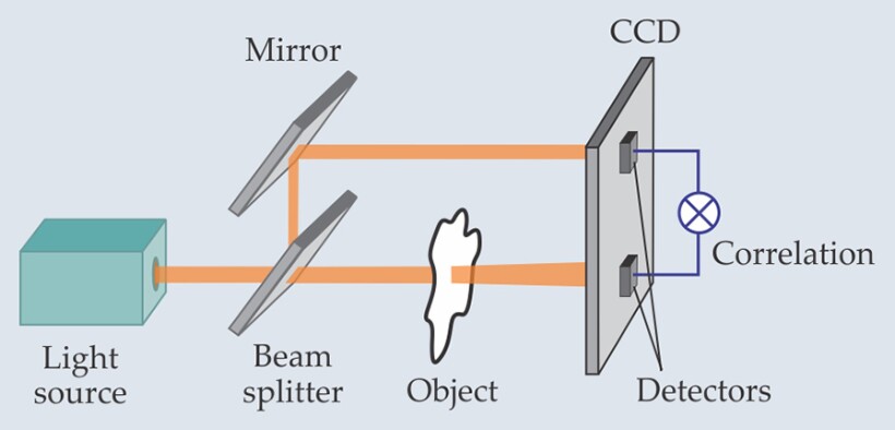
In its basic form, ghost imaging is a technique that indirectly produces a likeness of an object by combining the information from two light detectors—one that views the object and one that doesn’t. By splitting a speckled laser beam so that two beams strike spatially separated locations on a CCD camera screen and by placing an object in one of the beams, as shown here, researchers can calculate the intensity correlations between the two signals as the beams are scanned in synchrony and construct the object’s ghost image. Demonstrated in 2002 using visible light, the technique holds promise for remote atmospheric sensing because of its insensitivity to turbulence. Two research groups have now extended ghost imaging to the hard x-ray regime. Daniele Pelliccia (RMIT University in Australia) and his colleagues passed an x-ray beam produced by the European Synchrotron source through a diffracting silicon crystal grating oriented to transmit part of the undiffracted beam at a slight angle from the diffracted beam. An ultrafast camera placed 20 cm downstream of the beamsplitter detected both signals—one of which was partially blocked by the imaging target, a copper wire. Because the system worked with individual synchrotron pulses, retrieving the wire’s ghost image took more than just calculating intensity correlations; the group also had to filter out low-frequency artifacts associated with mechanical vibrations induced by the synchrotron pulse rate. By contrast, Hong Yu (Chinese Academy of Sciences) and colleagues periodically moved their test object—a gold film with five slits—into and out of a synchrotron’s x-ray beam to create their intensity-correlated signals—no beamsplitter or frequency filtering required. Turbulence is not a problem for x-ray imaging, but radiation exposure is. Pelliccia and colleagues speculate that one could pass a weak beam through fragile tissue but use its intensity correlations with a stronger beam that never touches the tissue to produce a low-noise microscopy image. (D. Pelliccia et al., Phys. Rev. Lett., in press; H. Yu et al., Phys. Rev. Lett., in press.)


