Seeing the softer side of nanoparticles
DOI: 10.1063/pt.urgj.mxfq

Nanoparticles come in many different shapes, including rods, triangles, and stars. Those shapes affect the way they interact with their environment. For example, they can change the way they are taken in by cells , an important consideration for drug delivery applications. The solvents used to engineer the shape of nanoparticles contain organic molecules that attach to the nanoparticle surface as ligands and influence their growth. But exactly how ligands exert that control is poorly understood. That’s because direct observation of ligand–nanoparticle interactions has been nearly impossible. Now Sara Bals, of the University of Antwerp in Belgium, and colleagues have developed a technique to bring the hard-to-see molecules into clearer view.

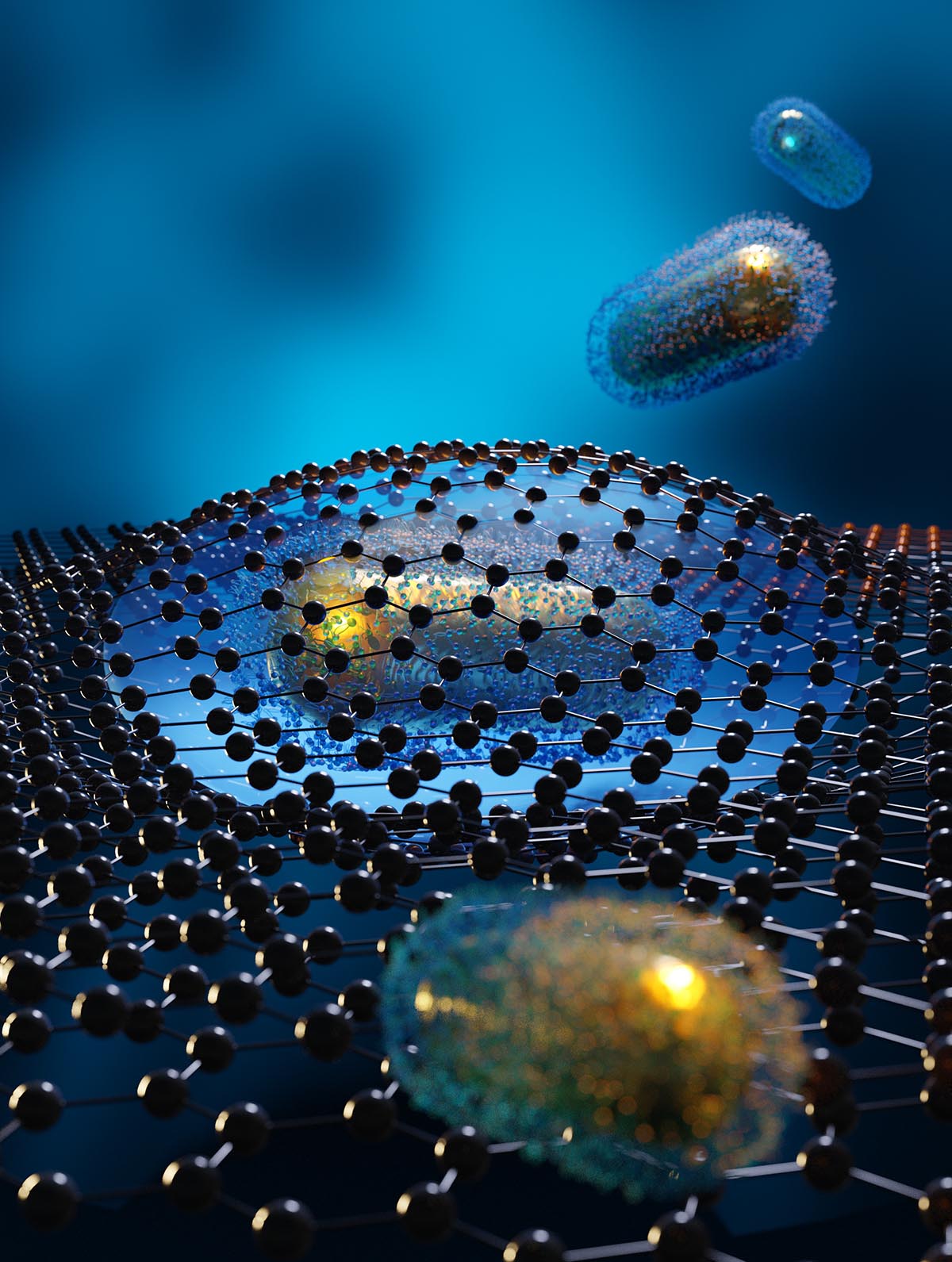
An illustration of a liquid cell trapped between two sheets of graphene, as part of a technique used to image the organic molecules that surround gold nanorods and influence their growth and behavior. Credit: Electron Microscopy for Materials Science, University of Antwerp
Electron microscopy, the typical method used to look at nanoparticles, is most sensitive to atoms with high atomic numbers, so carbon- and hydrogen-based ligands are hard to see. The grids normally used as a base for such imaging also contain a thick carbon layer that obscures the view of the ligands. And on top of that, the whole process is usually done in a UHV setting, which is nothing like the conditions in which ligands and nanoparticles are actually put to use. Bals and colleagues’ method uses pockets of liquid trapped between sheets of graphene to image nanoparticles and ligands in the liquid environment in which they do most of their work.
The use of ultrathin graphene as a base minimizes the background carbon signal so that the ligands can be imaged with higher contrast. Previous studies have used pillows of liquid between sheets of graphene to observe the growth and self-assembly of nanoparticles, but not to image ligands. Residues of polymers left behind from the typical graphene transfer procedure introduce contamination that obscures the view of the ligands. To develop a graphene transfer technique that left no polymer traces, the researchers used a polymer that they could remove by heating the graphene.
With the clean sheets of graphene in hand, the researchers looked at the ligands around gold nanorods in a solvent known to be a shape-directing agent—one that promotes the growth of the nanorod shape. What they saw surprised them. Previous scattering and spectroscopic imaging of gold nanorods in a dry setting had measured the thickness of the ligands, and from that thickness, it had been widely presumed that the ligands would form a bilayer around the rods. Though the new observations agreed with the previous measurements of thickness, the researchers saw no evidence of a bilayer.

Electron microscopy images of a gold nanorod in solution captured the moment when a ball of organic molecules moved toward the nanorod and attached to it. The interaction of organic molecules with nanoparticles in solution can dictate their size, shape, and interactions. Credit: A. Pedrazo-Tardajos et al., Nat. Chem. (2024)
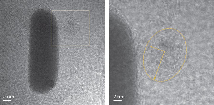
Rather, they observed in the solution a blob-like structure, known as a micelle, migrating toward and attaching to a nanorod, as shown in the figure. That brought into view the dynamic nature of ligands in solution. “Seeing the micelle was only possible because of the ideal conditions in the liquid cell,” says Nathalie Claes, a member of the research team. The technique offers a path forward for answering many open questions about how ligands shape and interact with nanoparticles. For example, how do ligands contribute to the self-assembly of nanoparticles into organized patterns? And how do ligands and micelles aid the growth of chiral nanoparticles from achiral seeds? (A. Pedrazo-Tardajos et al., Nat. Chem., 2024, doi:10.1038/s41557-024-01574-1 .)
This article was originally published online on 26 July 2024.
More about the authors
Laura Fattaruso, lfattaruso@aip.org


