Optical spectroscopy goes subnanometer
DOI: 10.1063/PT.3.2067
When photons interact with matter, they can scatter inelastically and gain or lose energy. Their frequency accordingly shifts by an amount that corresponds to a vibrational excitation. That Raman effect is the basis for a decades-old spectroscopic tool that identifies molecules by their unique vibrational fingerprints. Because the scattering cross section is low, the Raman signal is feeble. But it can be amplified more than a millionfold by, for example, placing the molecules in the tight space between a surface and the sharp metal tip of a scanning tunneling microscope (STM).
The simple act of shining light on the tip excites surface plasmons, collective electron-density oscillations in the metal. Those plasmons enhance the optical fields and localize them to a scale—typically tens of nanometers—far below the light’s diffraction limit. The sharper the tip, the greater the localization (see the article on near-field imaging by Lukas Novotny, Physics Today, July 2011, page 47 ). Using the technique, known as tip-enhanced Raman spectroscopy (TERS), researchers can map the topographic structure and vibrational spectra of molecules as a function of position (see the article by Katrin Kneipp, Physics Today, November 2007, page 40 ).
Zhenchao Dong and Jianguo Hou, both at the University of Science and Technology of China, and their colleagues have now implemented the first cryogenic (80 K) version of TERS in ultrahigh vacuum and improved the technique’s spatial resolution to less than 1 nm. 1 That’s nearly four times better than the previous state of the art. 2 As a demonstration, they imaged a single organic molecule, porphyrin, illustrated on page 15.

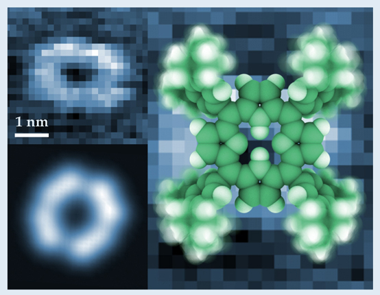
A single porphyrin molecule is illustrated (right) with its four lobes blurred to emphasize the molecule’s three dimensionality. A tip-enhanced Raman scattering image (top left) of a single molecule adsorbed on a silver surface produces the same ring structure that shows up in the simulated signal (bottom left).
In the experiment, both tip and surface were composed of silver, a metal thought to produce the strongest enhancements to a Raman signal. The coupling between the tip and surface in a light field drives the electrons back and forth and boosts the signal enhancement—particularly when they are driven at their natural resonance frequency.
The team illuminated the tip with green laser light and monitored the Raman-shifted red light backscattered from the porphyrin. Each of the scattered light’s Raman peaks signifies one of many vibrational modes. Subtle variations in the detected intensity of one of those modes (920 cm−1) produced the ring-shaped image (top left in the
Dong and colleagues also demonstrated how the molecule’s full set of vibrational modes is influenced by its orientation. As the bonding configurations between molecule and silver surface differed—with the porphyrin adsorbed flat onto a silver terrace, say, or at an angle on a silver step edge—so did the position of the Raman peaks. To see how much, the researchers positioned the tip over identical molecular features for side-by-side comparison of the spectra.
The system’s low temperature boosted the signal-to-noise ratio by suppressing the molecule’s diffusion and desorption, and its ultrahigh vacuum environment preserved a pristine substrate and stabilized the tip within a nanometer or so above a molecule. Those conditions, says Dong, allowed the team to precisely tune the plasmon resonance frequency to match that of the Raman emission for maximum enhancement.
Locating and controlling the plasmon frequency has traditionally been difficult. To find it, the researchers developed a clever approach: lower the tip to a molecule and raise the STM tunneling current until the porphyrin begins to glow; conveniently, the luminescence serves as a diagnostic for the plasmon resonance. And because the resonance depends not just on the tip’s composition but also on its size and shape, the researchers found they could sharpen the tip in situ, using an ion sputtering gun, and monitor the changing sample luminescence until its broad envelope overlapped both the incident laser frequency and specific Raman emissions.
When the resonances matched, the TERS signal rose. What’s more, it did so nonlinearly with incident laser power. The nonlinearity may suggest that some new, unconventional TERS mechanism is at play. “Unlike conventional TERS, in which the intensities of incident and Raman-shifted photons are linearly related, some kind of higher-order Raman process may confine the tip–sample interaction more tightly,” speculates Northwestern University’s Richard Van Duyne.
Details of the mechanism remain unclear. But the implications of pinning them down are broad: Catalysis, photochemistry, DNA sequencing, and protein folding, all imaged at the single-molecule scale, are just a few applications in a very long list.
References
1. R. Zhang et al., Nature 498, 82 (2013). https://doi.org/10.1038/nature12151
2. See, for example, R. Treffer et al., Biochem. Soc. Trans. 40, 609 (2012); https://doi.org/10.1042/BST20120033
T. Ichimura et al., Phys. Rev. Lett. 102, 186101 (2009).https://doi.org/10.1103/PhysRevLett.102.186101


