New technique makes brains transparent
DOI: 10.1063/PT.3.1997
The brain is a network: Its function depends critically on its connectivity. A neuron’s role in the brain depends far less on its spatial position than on how it connects to other neurons. And those connections, or synapses, are buried among a dense tangle of thin, highly branched axons and dendrites, the neuronal wires. Each just a micron or so thick, the wires form circuits that may span many millimeters or more. Understanding the brain means understanding its structure over many orders of magnitude in length.
For more than a century, neuroscientists have selectively stained small numbers of neurons in their entirety, so that their axons and dendrites, viewed through a light microscope, could be distinguished from those of their neighbors. More recently mice and other animals have been genetically engineered so that some or all of their brain cells synthesize proteins that fluoresce in various colors. 1 (See also Physics Today, December 2008, page 20 .)
As shown in figure 1a, though, brain tissue is opaque to light. To image individual neurons beneath the surface, researchers typically slice the brain into thin sections and image each one. Or, in a technique called serial block-face microscopy, they may alternately image and shave away the exposed surface of a solid block of tissue. 2 Either way, piecing together many cross-sectional images into a three-dimensional data set is a tedious process suitable only for small volumes of tissue.

Figure 1. An adult mouse brain (a) before and (b) after processing by the Clarity technique. (Adapted from ref.
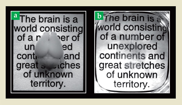
Now Stanford University researchers led by Karl Deisseroth have developed a technique, which they call Clarity, for making postmortem brains completely transparent, as shown in figure 1b, without destroying the fluorescent proteins and other molecules of interest. 3 Furthermore, the newly transparent brains are permeable to macromolecules, so additional fluorescent stains can subsequently be added. “This work may well be a game changer,” says Andreas Burkhalter of Washington University in St. Louis. “It promises to link structure to function at the level of neurons and synapses.”
Brain washing
The brain’s opacity is almost entirely due to lipid bilayers—the membranes that enclose cells, cell nuclei, and other organelles. But those membranes also give the tissue its structural stability. Deisseroth and colleagues’ strategy was to create a new way of holding the tissue together and then remove the lipids.
They began by infusing the brain with monomers of acrylamide and bisacrylamide (collectively known as hydrogel) and formaldehyde. The formaldehyde molecules attached to the brain’s proteins, nucleic acids, and some other molecules—but not to the lipids. The hydrogel monomers, in turn, attached to the formaldehyde. Incubating the brain at 37 °C polymerized the hydrogel. The result was a brain permeated with a solid polymer mesh that held protein molecules in place but allowed lipid molecules to move freely.
The lipids were removed from the brain by much the same process as a grease stain is removed from a shirt: Detergent micelles encapsulated the lipids, made them soluble in water, and allowed them to be washed away. But ordinary detergent moving through the brain by passive diffusion would take many months to do the job. Instead, the researchers used a detergent whose molecules carry an electric charge, drove the molecules through the tissue with an electric field, and cleared the brain of lipids in a mere five days.
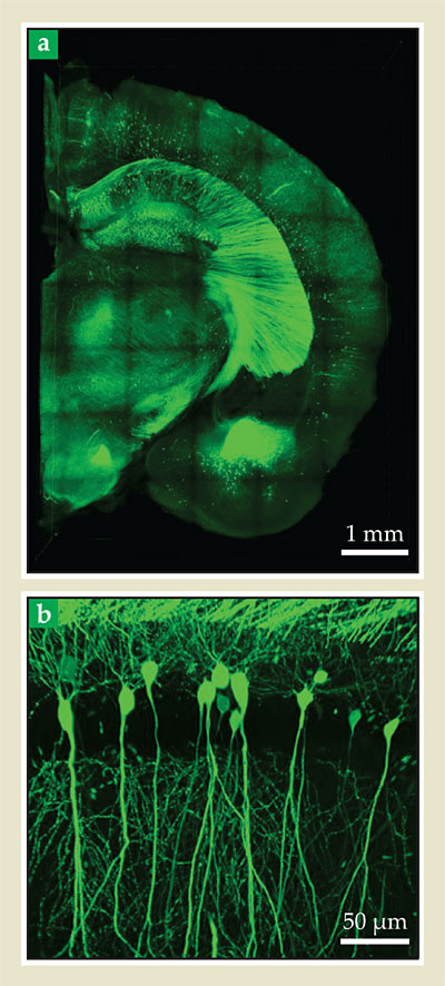
What was left of the brain was optically transparent. But were its proteins still there, still in place, and still able to fluoresce? The answer was yes on all counts. The images in figure 2, of a 1-mm-thick block of mouse brain genetically engineered so that some of its neurons express green fluorescent protein, show that Clarity preserves structure on both large and small scales. The researchers found that they could image up to 4 mm beneath the surface of the newly transparent brain with negligible loss of resolution—adult mouse brains, by contrast, are just 5–6 mm thick—and that improvements in their microscope optics might increase the imaging depth even more.

Figure 2. A portion of mouse brain, genetically engineered so that some of its neurons express green fluorescent protein, imaged to show both (a) large-scale structure and (b) individual neurons. (Adapted from ref.
Clarity is not the only way to make brain tissue transparent. For example, Ann-Shyn Chiang of National Tsing Hua University in Taiwan developed a cocktail of reagents, called FocusClear, that renders certain insect brains transparent almost immediately upon immersion. 4 Chiang and colleagues have used FocusClear to map portions of a cockroach brain; it’s less well suited to larger tissues. And, says Chiang, “We still don’t know what has been changed in the tissue by the FocusClear.”
Of mice and men
The Clarity technique also allows access to chemical information that’s not reflected in the distribution of genetically expressed fluorescent proteins. By infusing the transparent brain with fluorescent dyes bound to antibodies, which selectively latch onto a specific biomolecule of interest, Deisseroth and colleagues could image the distribution of any molecule for which an antibody can be designed. Notably, they can use that technique to fluorescently image preserved human brains, which for obvious reasons cannot be genetically engineered. The same ionic-detergent wash that removed the lipid membranes also removed the antibodies and thus prepared the brain for a new round of antibody staining.
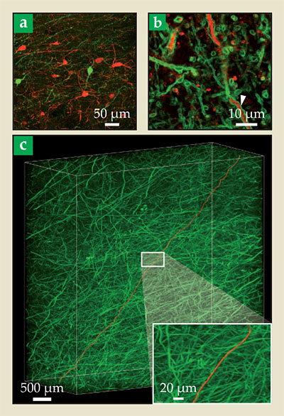
With the antibody technique, the researchers can fluorescently highlight whole neurons or parts of them, as shown in figure 3. They can also highlight synapses, by using antibodies that bind to molecules that are localized on the axonal and dendritic ends of the neuron connection.

Figure 3. Human brain tissue made transparent and imaged by antibody staining. (a) Red and green fluorescence signals indicate two proteins, parvalbumin and tyrosine hydroxylase, each expressed by a subset of whole neurons. (b) Here, the red signal is parvalbumin and the green is myelin basic protein, a substance contained in the electrically insulating sheaths that enclose the neurons’ axons. The white arrowhead indicates the myelin sheath around a parvalbumin-containing axon. (c) This three-dimensional rendering shows axons only. The red line (false color) shows a single axon traced across several millimeters. (Adapted from ref.
A complete map of even a portion of the human brain is still many years—maybe generations—away. The adult human brain contains some 100 billion neurons and perhaps a quadrillion synapses; it would be a daunting challenge just storing the brain’s connectivity map, let alone acquiring the map (for which automated image-tracing techniques are needed) or making sense of it. In the meantime, there are insights to be gained by studying brain connectivity on coarser scales—looking at connections between bundles of neurons rather than single cells—and on smaller scales. For example, Deisseroth and colleagues have shown that Clarity can reveal differences in the patterns of neuron connections in autistic and neurotypical brains.
References
1. G. Feng et al., Neuron 28, 41 (2000); https://doi.org/10.1016/S0896-6273(00)00084-2
J. Livet et al., Nature 450, 56 (2007). https://doi.org/10.1038/nature062932. W. Denk, H. Horstmann, PLoS Biol. 2, e329 (2004). https://doi.org/10.1371/journal.pbio.0020329
3. K. Chung et al., Nature (in press), https://doi.org/10.1038/nature12107 .
4. A.-S. Chiang et al., J. Comp. Neurol. 440, 1 (2001); https://doi.org/10.1002/cne.1365
Y.-C. Liu, A.-S. Chiang, Methods 30, 86 (2003). https://doi.org/10.1016/S1046-2023(03)00010-0
More about the authors
Johanna L. Miller, jmiller@aip.org


