Lauterbur and Mansfield Awarded Nobel Medicine Prize for Magnetic Resonance Imaging
DOI: 10.1063/1.1650215
This year’s Nobel Prize in Physiology or Medicine is shared by an American chemist and a British physicist “for their contributions concerning magnetic resonance imaging.” In the 1970s, Paul Lauterbur at SUNY Stony Brook and Peter Mansfield at the University of Nottingham in England set in motion the transformation of nuclear magnetic resonance (NMR) technology from a spectroscopic laboratory discipline to a clinical imaging technology that nowadays rivals, and in many ways surpasses, x-ray tomography in its ability to look inside the human body. (See the article by Felix Wehrli in Physics Today, June 1992, page 34 .)
Unlike x rays, isotope tracers, and endoscopes, MRI does not subject patients to ionizing radiation or other invasive risks. And by exploiting the variation of nuclear spin relaxation times and water concentrations, MRI can provide far greater contrast between different tissues and fluids than can x-ray absorption, which depends almost entirely on electron density. The relaxation times are parameters that characterize the decay rates of NMR signals.
From NMR to MRI
In 1946, Edward Purcell at Harvard and Felix Bloch at Stanford discovered nuclear magnetic resonance. They found that nuclei of nonzero spin aligned in a uniform magnetic field B can be resonantly reoriented to higher-energy Zeeman states by an RF field whose frequency ω precisely matches their Larmor precession frequency γB, where γ is the gyromagnetic ratio of the nuclear species. In more quantum mechanical language, the resonance condition is that the energy of an RF photon must equal the transition energy between Zeeman states. Bloch and Purcell shared the 1952 Nobel physics prize.
In 1971, physician Raymond Damadian at the Downstate Medical Center in Brooklyn reported anomalously long NMR relaxation times in excised mouse tumors. Later that year, Lauterbur, who was already known for his NMR studies of organic molecules with carbon-13, was watching Donald Hollis’s group at the Johns Hopkins Medical Center repeat and expand on Damadian’s experiment. “I began to wonder if there was a way to use NMR to localize tumors in a patient’s body,” he recalls. It soon occurred to Lauterbur that imposing small linear gradients on the uniform magnetic field of an NMR spectrometer might do the trick.
For the hydrogen nucleus—the spin-
In March 1973, Lauterbur published the first MRI image 1 (see figure 1). To produce this cross-sectional view of two 1-mm-diameter glass tubes of water in a bath of heavy water (D2O), Lauterbur used a commercial NMR spectrometer in an eccentric new way: Inevitable imperfections in the nominally uniform magnetic field of such instruments cause undesirable broadening of NMR spectral lines. Therefore the spectrometers were equipped with correcting coils that could produce weak field gradients to sharpen lines by compensating locally for field inhomogeneities. “I purposely cranked the correcting gradient all the way up in the wrong direction,” recalls Lauterbur. The changing intensity of the NMR signal as ω (or B) is swept then becomes a projection of the sample’s spatial distribution of hydrogen nuclei on the gradient axis. “And if I reoriented the sample or the gradient in enough different directions, I figured I could construct an image of the distribution of hydrogen nuclei.”

Figure 1. The first magnetic resonance image, published by Paul Lauterbur in 1973, was a cross section of two 1 -mm-diameter tubes of water in an NMR spectrometer. The curves in the left panel show the dependence of the NMR signal on the variable strength of the instrument’s uniform magnetic field (at constant radio frequency) when small linear field gradients are added in four different directions. They are, in effect, projections of the sample’s spatial distribution of hydrogen nuclei. Combining the four projections by a geometric algorithm yielded the two-dimensional image in the right panel.
(Reproduced from ref. 1.)

The left panel of figure 1 shows how the RF absorption signal of Lauterbur’s water-tube setup varied with changing B for fixed ω and four different orientations of the gradient relative to the sample. From those four projections, he constructed the two-dimensional image (right panel) by devising a back-projection algorithm similar to those just starting to be used at the time for computer-assisted x-ray tomography.
In a variation of the experiment that addressed his initial motivation, Lauterbur dissolved a trace amount of a paramagnetic salt in the water of one of the two tubes to shorten T 1 the characteristic relaxation time for flipped nuclear spins to regain their equilibrium alignments. At sufficiently high RF power, the NMR signals are weakened by saturation; once a spin has been flipped, it won’t flip again before it has relaxed to its lower-energy alignment. Finding that such saturation is significantly inhibited in the doped water with the higher T 1 Lauterbur wrote that “a possible application of considerable interest would be the in vivo study of malignant tumors.” 1
Magnetic field gradients had been used previously in NMR to provide some spatial information in one dimension: Erwin Hahn at the University of Illinois, and Purcell and Herman Carr at Harvard, had exploited field gradients in the 1950s to study the flow and diffusion of fluids. At Cornell in 1972, David Lee, Douglas Osheroff, and Robert Richardson used NMR with a gradient to localize different phases of helium-3 in their discovery of its superfluidity But Lauterbur was the first to undertake actual imaging.
Noting that his new technique was a coupling of two fields—magnetostatic and RF—by the object being imaged, Lauterbur suggested the name zeugmatography, after the Greek word ζευγµα, which means a yoking together. Thirty years later, although the technique has flourished beyond anything he imagined in those early days, the name hasn’t stuck. Also vanished from the now universal appellation magnetic resonance imaging is the descriptive but impolitic adjective nuclear.
“A whole complex of applications and refinements had to be explored before clinicians and manufacturers became seriously interested in the early 1980s,” says Lauterbur. For the rest of the 1970s, he and various coworkers at Stony Brook investigated the exploitation of fluid flow, relaxation times, chemical shifts of the hydrogen spin resonance in different molecular environments, and the mapping of nuclei other than hydrogen. 2 And there was the ongoing quest for instruments large and fast enough to accommodate patients, and for stronger and more uniform magnetic fields.
The quest for speed
In the summer of 1973, not yet knowing of Lauterbur’s recently published paper, Mansfield and Peter Grannell, his postdoc at Nottingham, were also exploiting magnetic field gradients to do NMR imaging of a different kind. 3
Mansfield, a solid-state physicist, had been using NMR to study inorganic crystals and dilute metal alloys. A persistent problem of doing NMR studies in solids was the broadening of spectral lines by magnetic dipole interactions between adjacent nuclei. Having developed sophisticated sequences of RF pulses to combat this kind of broadening, Mansfield thought about how one might use the narrowed NMR lines together with a linear magnetic field gradient to examine the spatial structure of crystals.
The finer the spatial resolution one wants, the steeper must be the gradient. Unfortunately, resolving the angstrom-scale structure of a crystal would require impossible gradients of order 108 G/cm. So Mansfield and Grannell set out to test their idea on a more macroscopic scale, with simple artificial crystals—arrays of thin camphor planes spaced at intervals of about 1 mm—in an NMR spectrometer to which they added a field gradient of 0.8 G/cm normal to the planes. (The carbon and oxygen nuclei in hydrogen-rich camphor, and in biological tissue, are spinless, except for some minority isotopes.)
A faster, pulsed alternative to looking for NMR absorption by sweeping B or ω in a continuous-wave RF field is to subject the sample to a short, intensive “90° pulse” that flips all the proton spins at once into the plane perpendicular to B. For a time, the flipped protons precess coherently in the magnetic field, producing a weak free-induction-decay (FID) radio frequency signal that can be monitored as it decays by relaxation and increasing incoherence. The Fourier transform reproduces the continuous-wave NMR spectrum.
Because the precession frequency increases linearly with position along an imposed uniform gradient, the Fourier transform of the FID signal yields the projection of the hydrogen density distribution on the gradient direction. Performing this experiment with several of their artificial crystals, Mansfield and Grannell found that the Fourier transforms clearly marked the positions of the individual planes with submillimeter resolution.
“That was, I believe, the first demonstration of NMR diffraction in a solid,” says Mansfield. But in awarding him the Nobel Prize, Sweden’s Karolinska Institute emphasized his contributions throughout the 1970s to speeding up the acquisition and display of images, which was essential to the development of MRI as a useful clinical technique.
In 1974–75, Mansfield and coworkers developed techniques for scanning samples rapidly by selective RF excitation of thin planar and strip slices in a sample subjected to a sequence of gradients in different directions. A planar slice is selected by imposing on the entire sample a field gradient normal to the slice and subjecting the sample to an RF pulse whose frequency content is tailored to saturate all the proton spins except those in the desired slice. Then one quickly switches to an orthogonal gradient and, with another appropriately tailored RF pulse, excites only a selected strip (perpendicular to that second gradient) in the unsaturated slice.
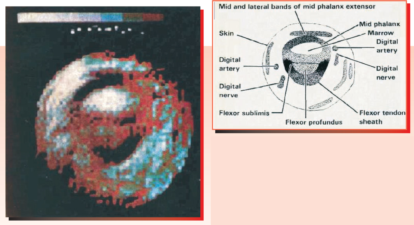
In 1976, this “line-scan” technique produced the first MRI image of a living human body part 4 (see figure 2). The appendage thus immortalized was a finger of Mansfield’s graduate student Andrew Maudsley. “When I first showed the color-coded image at a meeting in London,” recalls Mansfield, “people were astonished at the anatomic detail.” The cross-sectional image showed individual muscles, tendons, bone marrow, arteries, and even nerve fibers. Much of the contrast is due to the T 1 sensitivity of the image, achieved by repeated excitation of the same strip with different subsecond time delays.

Figure 2. The finger of Andrew Maudsley, Peter Mansfield’s student in 1976, provided the first human in-vivo MRI image. The color coding indicates the intensity of different pixels. The labeled tracing of the cross-sectional image shows the impressive anatomical detail already revealed by MRI in those early days.
(Reproduced from ref. 4.)
Mansfield’s introduction of echo planar imaging (EPI) in 1977 took MRI to an entirely new realm of speed. 5 EPI nowadays makes it possible to repeatedly image an entire brain many times a second. Such speed lets physicians pinpoint and evaluate brain tumors by following the leakage of blood through lesions.
EPI exploits the spin-echo effect discovered by Hahn in 1949. After a 90° RF pulse in a uniform magnetic field, the FID signal dies out exponentially as precessions lose their phase coherence due to interactions between neighboring spins. T 2, the characteristic time for this “spin–spin relaxation,” is shorter than T 1 Hahn discovered that a second RF pulse, delivered before decoherence is complete, generates an “echo” signal that resembles the waning original FID signal. The second pulse creates the echo by initiating a time reversal of the growing phase decoherence of the spin precessions.
Mansfield proposed that one could use spin echoes to image an entire plane of a sample, or even its full volume, with just a single 90° excitation pulse. In the planar case, one field gradient in the selectively excited plane would be repeatedly reversed at intervals much shorter than T 2, thus regenerating spin echoes much as repeated RF pulses would. The time discreteness introduced by the rapid switching, he pointed out, together with a steady gradient in the orthogonal direction in the plane, can be made to impose on the continuous spin distribution a quasi-discrete structure of lattice points, each with its own unique precession-frequency signature.
EPI is supplanting functional positron-emission tomography for the real-time study of brain function in cognitive science. Beyond its sensitivity to blood flow, MRI, unlike PET, can distinguish between oxygenated and de-oxygenated hemoglobin by the latter’s strong paramagnetism. More quotidian tasks like imaging a torn ligament don’t require the ultra-fast capabilities of EPI and its exacting hardware requirements. Ordinary clinical MRI relies more on 2-D Fourier-transform techniques introduced in 1975 by Richard Ernst at ETH Zürich. Ernst won the 1991 chemistry Nobel Prize for his contributions to NMR spectroscopy.
The laureates
Lauterbur was born in 1929 in Sidney, Ohio. After his bachelor’s degree in chemistry from the Case Institute of Technology, he started as a research assistant at the Mellon Institute in Pittsburgh. In 1962, while still a full-time researcher at Mellon, Lauterbur received his PhD at the University of Pittsburgh. “I did carbon-13 NMR, but I had no real thesis adviser,” he recalls.
The following year, Lauterbur joined the Stony Brook chemistry faculty. In 1985 he left Stony Brook for the University of Illinois at Urbana, where he is a professor of chemistry at the Center for Advanced Study. “I recently stopped doing MRI,” says Lauterbur, “to get back to my roots as a chemist after a 30-year detour.” His current interest is prebiotic evolution.
Mansfield is a Londoner, born in 1933. His scientific career had an atypical start. In the turmoil of returning home in 1944 after having been evacuated from London during the Blitz, he did poorly in the secondary-school entrance exam and ended up in a high school that offered no college-preparatory program. So he left school at age 15 to become an apprentice typesetter.
But the German V-1 and V-2 missiles that began pounding London in the summer of 1944 piqued young Mansfield’s interest in rocketry and explosives. At age 17 he got a job in a government rocket laboratory, where he worked until 1956. Then, having gone to night school and passed the required exams, he entered Queen Mary College in London, where he did his undergraduate and graduate work in physics. He got his PhD under Jack Powells in 1962, doing NMR studies of solids. He came to Nottingham in 1964.
Mansfield was knighted in 1993. A year later he retired from the faculty, but he still works to improve MRI. At the moment he’s seeking ways of reducing the horrendous noise to which patients are subjected as gradient coils are vibrated by the rapid switching of currents in the scanner’s multitesla field.
Three days after the Karolinska Institute announced this year’s prize for MRI, full-page ads appeared in several major newspapers in the US and Sweden, decrying “the shameful wrong that must be righted,” that is, the exclusion of Damadian from the prize. He is president of the Fonar Corporation, a firm that manufactures conventional MRI instruments.
Lauterbur has been the object of Damadian’s particular ire ever since he omitted Damadian’s 1971 paper on anomalous relaxation times in tumors from the reference list of his first imaging paper. 1 “I chose, instead, to reference the only published paper on tumor NMR in a living animal,” explains Lauterbur. In 1974, Damadian was awarded a patent for an NMR instrument designed to scan the body for cancerous growths. The design did not involve the linear field gradients that are the basis of all modern MRI instruments, including those sold by Fonar.

Lauterbur
BILL WEIGAND

Mansfield
PAGE ON
References
1. P. C. Lauterbur, Nature 242, 190 (1973) https://doi.org/10.1038/242190a0 .
2. P. Bendel, C. M. Lai, P. C. Lauterbur, J. Magn. Res. 38, 343 (1980) https://doi.org/10.1016/0022-2364(80)90285-1 .
3. P. Mansfield, P. K. Grannell, J. Phys. C 6, L422 (1973) https://doi.org/10.1088/0022-3719/6/22/007 .
4. P. Mansfield, A. A. Maudsley, Brit. J. Radiol. 50, 188 (1977) https://doi.org/10.1259/0007-1285-50-591-188 .
5. P. Mansfield, J. Phys. C 10, L55 (1977) https://doi.org/10.1088/0022-3719/10/3/004 .


