Ghost imaging delivers high resolution with a limited x-ray dose

X-ray imaging with high spatial resolution is widely used in both scientific and clinical applications to look inside samples noninvasively. But although it allows users to quickly assess a fractured bone or the atomic structure of a crystal, such imaging can cause significant cellular damage to biological samples when they are extensively subjected to its ionizing radiation. Now Tiqiao Xiao of the Chinese Academy of Sciences in Shanghai and colleagues have adapted a technique called ghost imaging, most of whose x rays never interact with the sample. Scanning nonbiological samples, the researchers achieved resolution on par with what’s needed for biomedical applications while exposing samples to a fraction of the x-ray photons typically required in radiology. 1
Figure 1.

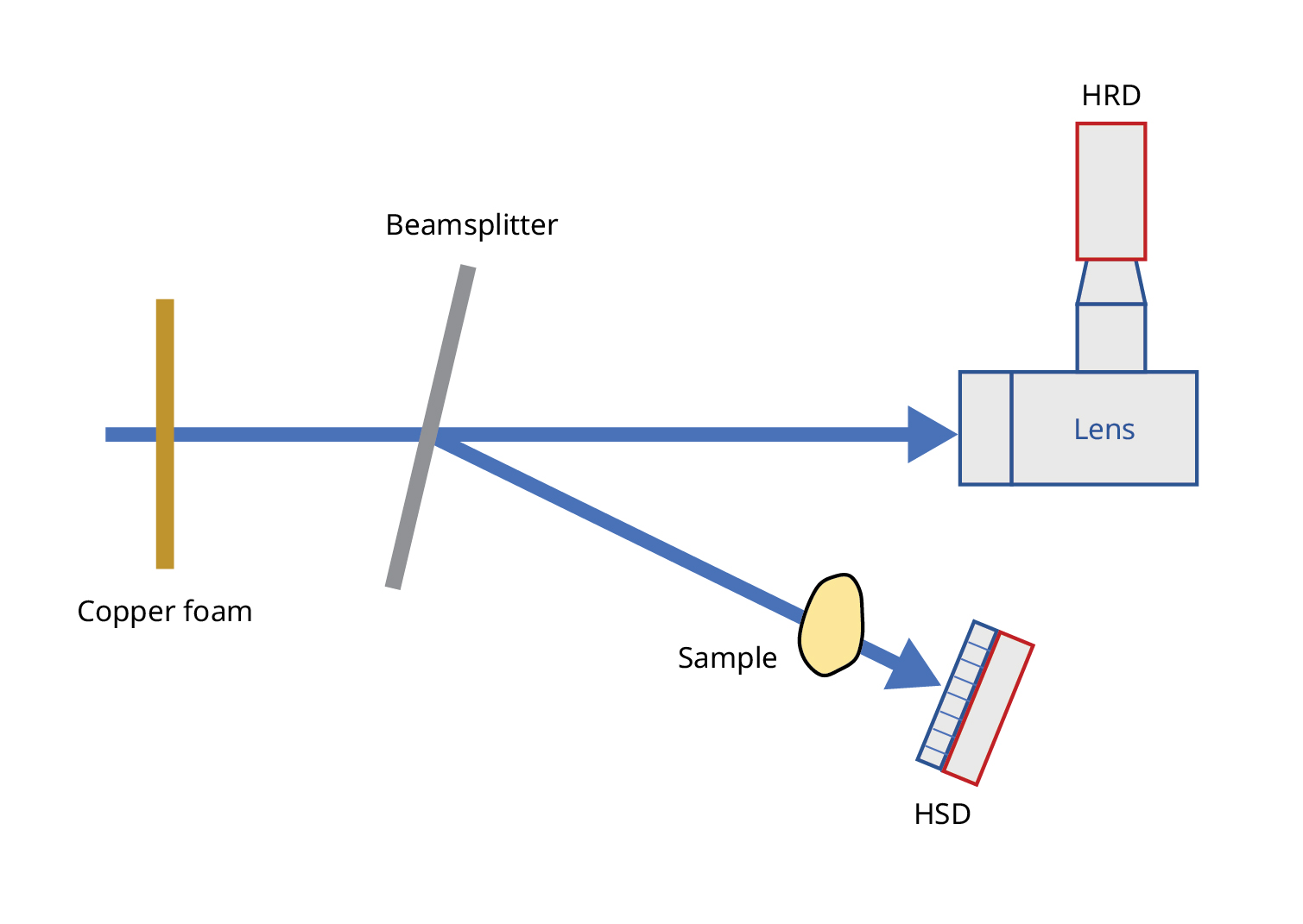
The x-ray imaging technique implemented by Tiqiao Xiao and colleagues uses copper foam to imprint a beam with a speckle pattern before splitting it into two components: a ghost, or reference, beam that never directly interacts with a sample and an object beam that does. By correlating measurements taken by specialized—high spatial resolution (HRD) and high photon sensitivity (HSD)—detectors at the end of the beams’ paths, the team can reconstruct an image.
(Image adapted from ref. 1 .)

Ghost imaging was first experimentally demonstrated in 2016. As shown in figure 1 , in Xiao and colleagues’ setup, an x-ray beam is imprinted with a unique pattern before it’s split to create a ghost, or reference, beam that never directly interacts with a sample and an object beam that does. Measurements of the spatial features of the reference beam are matched with measurements of photon intensities of the object beam to gradually reconstruct an image of the sample.
A major challenge is that unlike radiological imaging, ghost imaging requires multiple scans of the sample, which can add to the total photon count even if the per-scan exposure is relatively low. To achieve high resolution while minimizing the number of scans, Xiao and colleagues installed a crystal beamsplitter and detectors tailored to each of the split beams. At the Shanghai Synchrotron Radiation Facility, the researchers passed an x-ray beam through a 300-μm-thick copper foam sheet to encode it with a speckle pattern and then split the beam with the crystal. The reference beam traveled directly to a high spatial resolution detector, while the object beam passed through a sample before reaching a detector with high photon sensitivity.
Figure 2.

A circuit board is imaged with both a new x-ray ghost imaging technique (top) and conventional x-ray imaging (bottom). The new method required less than 0.5% of the photons of the conventional method to achieve the same resolution.
(Images courtesy of Tiqiao Xiao, Shanghai Advanced Research Institute, Chinese Academy of Sciences.)

To compare the image quality of the new method, dubbed ultra-low-light megapixel x-ray ghost imaging (UMXGI), with that of traditional x-ray imaging, the researchers used both techniques to reproduce a circuit board. They looked at the contrast-to-noise ratio—a measure of how much an object stands out from the background noise in an image—and the number of photons needed to visualize certain microstructures. The images are shown in figure 2 . The team consistently found that UMXGI achieved the same image quality as the traditional method with 0.48% of the number of photons interacting with the sample compared with traditional radiological imaging.
UMXGI is not ready to be used on biological samples because they generally have less predictable structures and require a higher contrast-to-noise ratio than was achieved by the recent experiment. The next steps toward that milestone, Xiao says, include transitioning to a laboratory-scale x-ray source, like an x-ray tube. The team also plans to investigate the use of advanced image-reconstruction algorithms to improve overall image quality.
Reference
1. C. Zhao et al., “Ultra-low-light megapixel X-ray ghost imaging ,” Optica 13, 1387 (2026).