Coulomb-explosion imaging tackles an 11-atom molecule
DOI: 10.1063/PT.3.4994
Sometimes the only way to get a good look at something is to destroy it. When archaeologists excavate a site, they forever disrupt the context of how the artifacts they find were arranged in the ground. For gas-phase chemists, the molecules of interest are so small and move so fast that they can’t be directly imaged—unless they’re blown to bits.
X-ray diffraction and electron diffraction, cousins of the similar but less destructive crystallography techniques used for studying ordered solid samples, have found success in resolving the structures of isolated biomolecules and viruses. (See Physics Today, April 2011, page 13 .) When it strikes a single molecule, the x-ray or electron pulse breaks bonds and destroys the specimen. Even so, a small fraction of the photons or electrons scatter elastically, just as they do in a crystal. From those particles’ diffraction pattern, researchers can extract structural information.
A complementary technique, Coulomb-explosion imaging, is especially well suited to studying smaller molecules in detail. Like diffractive imaging, it involves blasting a molecule with a pulse, usually of photons, that’s powerful enough to expel the molecule’s electrons, break its chemical bonds, and drive the now positively charged atoms to repel one another. But rather than studying the scattered photons, Coulomb-explosion imagers collect the charged fragments of the molecule itself. By measuring the fragments’ momentum and modeling the trajectories the ions followed as they flew apart, researchers can deduce the atoms’ starting positions: the molecular structure.
Coulomb-explosion imaging has revealed exotic molecules that are too weakly bound to study spectroscopically (see Physics Today, July 2015, page 10 ) and reaction pathways that had only ever been observed indirectly (see the Quick Study by Tomoyuki Endo, Chen Qu, and Heide Ibrahim, Physics Today, July 2021, page 62 ). But despite those successes, researchers have limited use of the technique—at least in its purest form—to molecules with approximately five or fewer atoms.
Two assumptions lay behind that presumed restriction. Getting a good Coulomb-explosion image requires placing a positive charge on every atom in a molecule, so that all the pair-wise atomic interactions are dominated by Coulomb repulsion rather than electronic attraction. Researchers assumed that it wouldn’t be possible to simultaneously ionize every atom in a large molecule. They also assumed that they could reconstruct a molecule’s structure only if they detected all of its ionic fragments—a daunting task for larger molecules when the detection efficiency for each ion hovers around 60%.
Now, working at the European X-Ray Free-Electron Laser, or EuXFEL, Rebecca Boll (seen in figure
Figure 1. Rebecca Boll works on the Small Quantum Systems instrument at the European X-Ray Free-Electron Laser. Wrapped in aluminum foil is the reaction microscope, which captures the charged fragments of molecules blown up by the powerful x-ray pulses. The foil insulates the equipment while the researchers bake out the residual gases. (Courtesy of European XFEL/Jan Hosan.) Figure 2. The 11-atom molecule 2-iodopyridine is laid bare by Coulomb-explosion imaging. (a) The molecular structure, which was already known, centers on a rigid, planar ring of carbon and nitrogen atoms. (b) When x-ray ionization breaks the chemical bonds and sends the atoms flying apart, the six ring atoms are readily distinguished by their momentum. (c) So are the four hydrogen atoms, the lightest and most challenging atoms to detect. (Adapted from ref. 


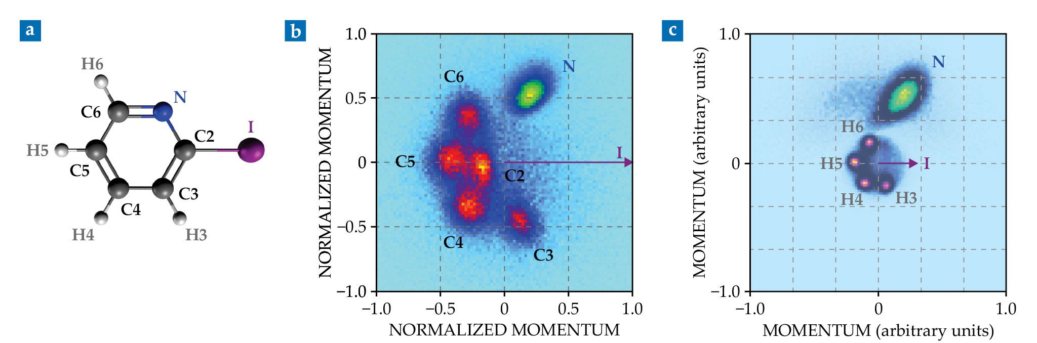
And even when the researchers detected as few as three of the atoms from any given molecule, the momentum distributions built up from repeated Coulomb explosions were crisp enough to distinguish all five of the molecule’s carbon atoms (as seen in figure
Unexpected clarity
The researchers didn’t set out to overturn the assumptions of Coulomb-explosion imaging. “If we’d said in our proposal that we wanted to image every individual atom in these molecules, we would have been laughed out of the room,” says Boll. Rather, the project started with the more modest aim of exploring molecular ionization dynamics under the influence of XFEL pulses.
Before the EuXFEL came on line in 2017, Boll and Santra were part of a team using SLAC’s Linac Coherent Light Source (LCLS), also an XFEL, to study ionization of iodomethane (CH3I) and iodobenzene (similar to 2-iodopyridine, but with an all-carbon hexagonal ring). 2 They noticed that the XFEL pulses, thanks to multiphoton absorption, removed an astonishing number of electrons from both molecules. Absorption was concentrated on the iodine atom, which had the largest absorption cross section in the molecule, and produced charge states as high as I47+. That’s no small feat when the atom has only 53 electrons to begin with.
The charge seemed to spread efficiently from the I atom to other parts of the molecule. In iodobenzene, the researchers saw carbon atoms as highly charged as C4+. But they didn’t know which C atoms they were seeing—the ones adjacent to the I atom or the ones on the opposite side of the ring. And the LCLS experiment wasn’t equipped to help figure it out.
Boll and Santra didn’t think that Coulomb-explosion imaging could distinguish two atoms of the same element in the same molecule—at least, not without detecting every atom in the molecule, which for a structure as large as iodobenzene would be nearly impossible. But the technique can easily distinguish atoms of different elements because of their different masses.
The original plan for the new EuXFEL experiment, then, was to replace one C atom at a time with a nitrogen atom. If the N atom in 4-iodopyridine (on the opposite side of the ring as the I atom) ended up just as highly charged as the one in 2-iodopyridine, it would follow that the charge is readily redistributed around the ring.
The researchers expected the pattern formed by all the other atoms to be a featureless blob. But to their surprise, when they plotted the C atoms’ momentum, the data formed five distinct bunches, one for each position around the ring. The four hydrogen atoms showed an even cleaner bunching. So even if the researchers detect only one of the C or H atoms from a given Coulomb explosion, they can tell which one it is just by measuring its momentum.
What a coincidence
The 2-iodopyridine molecules tumble randomly through space on their way to their destruction by the XFEL beam. The distributions in figure
The reorientation is possible because the researchers did what’s called coincidence mapping: They made their beam of 2-iodopyridine so dilute that each x-ray pulse interacted with at most one molecule. Only under those conditions could they be sure that all the ions detected in a single shot came from the same parent molecule. The data in figure
Coincidence-mapping experiments can take a long time to build up a meaningful data set. The EuXFEL’s high repetition rate, up to 570 pulses per second for these experiments, helps. Without access to such a high-powered facility, other research groups have explored an alternative technique called covariance mapping: simultaneously ionizing many molecules (usually after physically aligning them with polarized light) and using sophisticated data analysis techniques to make up for the fact that they can’t definitively trace any two atomic fragments to the same parent molecule.
Covariance mapping has yielded some structural, and even dynamical, information for molecules with more than 20 atoms. 3 “But those experiments are always losing some information,” says Jahnke. “For example, if you’re looking at an ensemble of molecules doing some dynamical process, those molecules aren’t all doing the same thing.” Coincidence mapping, although time-consuming and experimentally challenging, yields that molecule-by-molecule information.
Toward molecular movies
In their theoretical analysis, Santra and Schäfer didn’t focus on determining bond lengths, bond angles, or other structural details. The equilibrium structure of 2-iodopyridine, like those of most similarly sized molecules, is already known. Instead, starting from that established structure, the theorists simulated the Coulomb-explosion process and showed that it yields momentum distributions much like the ones that were observed. If necessary, they could iteratively adjust the starting configuration to converge on an unknown molecular structure.
Of greater interest than equilibrium structures, though, are the insights that Coulomb-explosion imaging can offer for molecules in motion: An ultrashort laser pulse initiates a chemical reaction, and the XFEL pulse instigates a Coulomb explosion tens or hundreds of femtoseconds later. Most of what researchers know about what happens during a reaction they learn indirectly, either by studying the reaction products (see, for example, Physics Today, February 2019, page 14 ) or by spectroscopically probing the molecules midreaction (see Physics Today, December 1999, page 19 ). Coulomb-explosion imaging provides a more direct look.
One barrier to studying the dynamics of 11-atom molecules is figuring out how best to visualize all the data. The two-dimensional momentum plots in figure
“X-ray diffraction and electron diffraction can resolve molecular structures,” says Santra, “but they’re purely three-dimensional. They can’t map out the whole molecular phase space, which is our goal. With Coulomb-explosion imaging, we’re still not detecting everything, but we can get so much more information than traditional techniques provide.”
References
1. R. Boll et al., Nat. Phys. (2022), doi:https://doi.org/10.1038/s41567-022-01507-0 .
2. B. Erk et al., Science 345, 288 (2014); https://doi.org/10.1126/science.1253607
A. Rudenko et al., Nature 546, 129 (2017). https://doi.org/10.1038/nature223733. C. Vallance, D. Heathcote, J. W. L. Lee, J. Phys. Chem. A 125, 1117 (2021); https://doi.org/10.1021/acs.jpca.0c10038
C. A. Schouder et al., Annu. Rev. Phys. Chem. 73, 323 (2022), doi:https://doi.org/10.1146/annurev-physchem-090419-053627 .
More about the authors
Johanna L. Miller, jmiller@aip.org


