Viruses in 3D
DOI: 10.1063/1.3141951
X-ray crystallography and cryoelectron microscopy have long confirmed Francis Crick and James Watson’s assertion that the protective protein shell, or capsid, of many spherical viruses would have icosahedral symmetry (see Physics Today, January 2008, page 42 , and December 2004, page 27 ). Most virus genomes are encoded in either single-stranded RNA of double-stranded DNA. One group of viruses, whose hosts range from bacteria to humans, has double-stranded RNA. Most of the dsRNA viruses share a common property not found in other viruses: Their icosahedral capsids consist of 120 subunit proteins arranged in 60 pairs, or dimers.
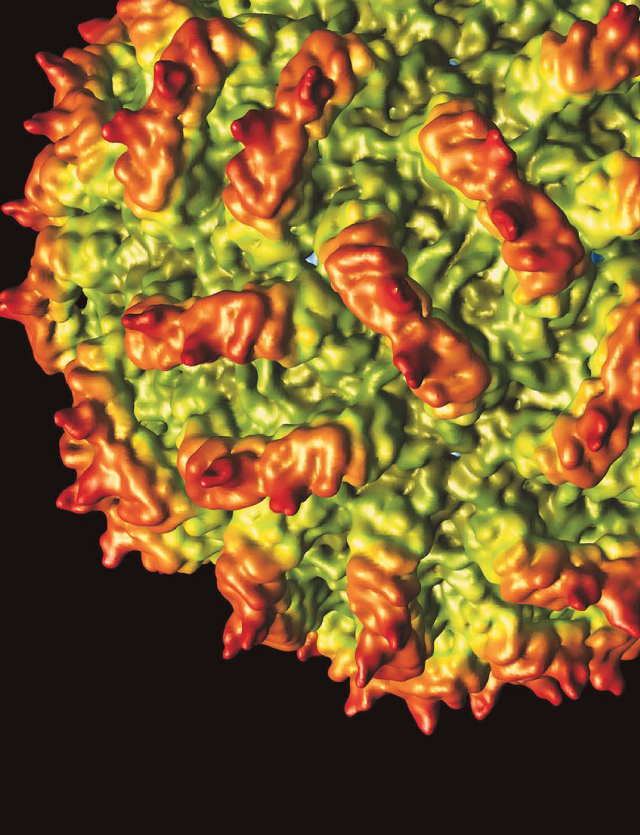
New work has determined the three-dimensional capsid structure of two members of the family Partitiviridae, one of the last dsRNA families to have its structure solved. Shown here is the Penicillium stoloniferum virus S, resolved to 7.3 Å and colored by radius. The 3D reconstruction was assembled from 14 246 individual particle images obtained with cryoEM. The 35-nm-diameter capsid is marked by 60 protruding arches, each formed by a protein dimer. A subtly different arched structure was found for the related P. stoloniferum virus F and resolved to 3.3 Å by x-ray crystallography. (W. F. Ochoa et al. Structure 16 , 776, 2008 http://dx.doi.org/10.1016/j.str.2008.02.014 ; J. Pan et al. Proc. Natl. Acad. Sci. USA 106 , 4225, 2009 http://dx.doi.org/10.1073/pnas.0812071106 .)
To submit candidate images for Back Scatter, visit http://www.physicstoday.org/backscatter.html .

Image: W. F. Ochoa, UC San Diego; source: San Diego Supercomputer Center, UCSD.


