A mouse-cortex snapshot at subcellular resolution


Neuroscientists have long known that blood flow to a particular area of the brain increases when neurons in that section are activated. The phenomenon, termed neurovascular coupling, enables the imaging of brain function on a local level through use of techniques such as functional MRI. But a broader understanding of the spatial and temporal features of neurovascular coupling at the cellular level has eluded researchers: It has proven difficult to develop an imaging technique with both the necessary field of view and resolution to capture brain activity on a cortex-wide level.
To solve that problem, a team led by Chengbo Liu at the Shenzhen Institutes of Advanced Technology in China has constructed a new type of hybrid microscope that integrates two imaging techniques: confocal fluorescence microscopy and photoacoustic microscopy. The first captures fluorescent calcium signals that are emitted during neuronal activity; the second, based on the photoacoustic effect—when sound waves form in a material after it has absorbed light—exploits the optical absorption of hemoglobin.
The microscope consists of an array of eight miniature high-frequency transducers, which extend the field of view for both microscopies without any loss of sensitivity in photoacoustic microscopy. The transducer array is coupled with a custom-built, 16-facet polygonal mirror to enable real-time imaging.
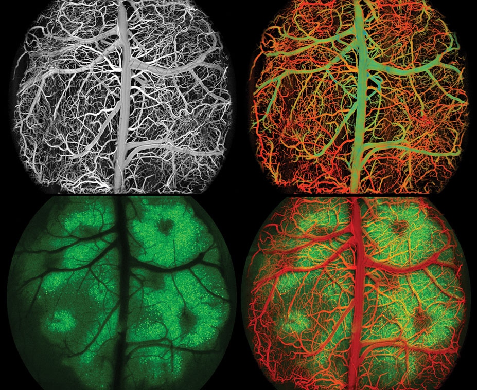
The team demonstrated the combined technique’s high resolution and field of view by imaging the neurons and blood flow in the dorsal cortices of mice that were in the resting state, under anesthesia, or in an induced epileptic seizure. These images from a resting mouse’s brain show a map of blood-vessel intensity (top left); a map of blood-oxygen saturation, in which red shading indicates higher oxygen levels (top right); a fluorescence-intensity map (bottom left); and the merged final image output, in which the blood vessels have been colorized in red for clarity (bottom right).
Liu and his team see the technique helping researchers study neurological disorders associated with aberrant neurovascular coupling, such as seizure disorders, strokes, and Alzheimer’s disease. They also envision miniaturized versions of the hybrid microscope allowing neurosurgeons to see brain oxygenation and activity in real time. (L. Liu et al., Sci. Adv. 11, eadw5275, 2025 ; images courtesy of Chengbo Liu laser-induced ultrasound lab, Shenzhen Institutes of Advanced Technology.)


