Hearing links
DOI: 10.1063/1.2800076
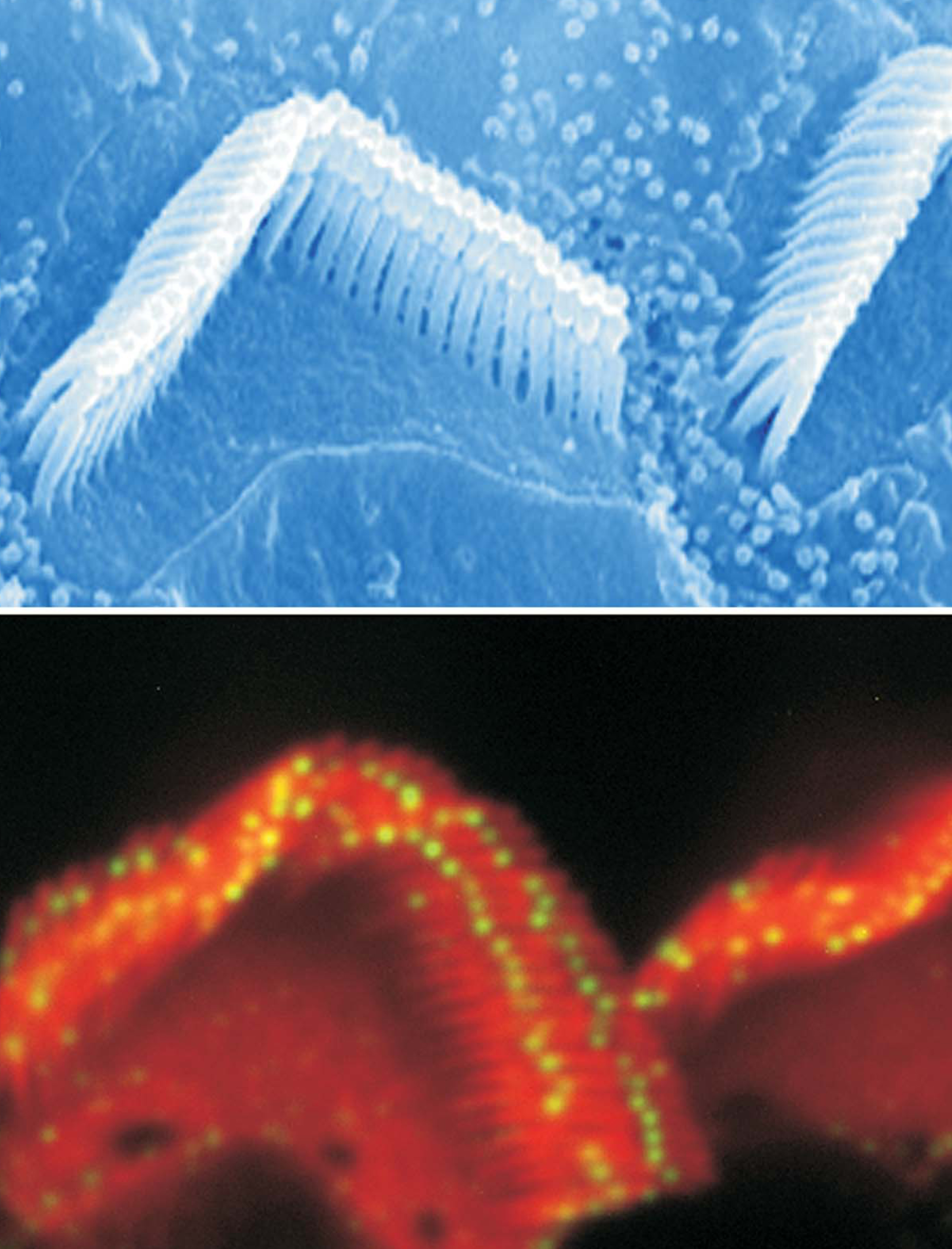
Hair cells in the inner ear are among the most sensitive mechanical transducers in the human body. The key players in the conversion of sound waves and motion into the electrical impulses that underlie our senses of hearing and balance are bundles of bristly stereocilia that protrude from the top of the hair cells. As seen in this scanning electron micrograph (top) of rodent hair cells, the stereocilia are arranged in rows, with each row taller than the one in front of it. Connecting the tops of stereocilia in adjacent rows are filaments called tip links. In addition to ensuring that the entire bundle moves together, the tip links are thought to be responsible for the mechanoelectrical transduction: As the stereocilia get pushed toward the taller rows, the tip links stretch and open ion channels that generate an electrical response; movement toward the shorter rows inhibits the opening of the channels. (For more about hair cells, see the article by A. J. Hudspeth and Vladislav Markin in Physics Today, February 1994, page 22 .)
New work by Bechara Kachar (National Institute on Deafness and Other Communication Disorders), Ulrich Müller (Scripps Research Institute and the Institute for Childhood and Neglected Diseases in La Jolla, California), and their coworkers has shown that two members of the cadherin family of transmembrane proteins constitute the tip links. Cadherin 23, tagged with a green fluorescent antibody in the fluorescence microscopy image (bottom), is found at the tall end of the tip link.
Protocadherin 15 is found at the short end, and the tails of the two proteins unite and adhere. Knowing the molecular composition of tip links, say the researchers, will provide the basis for further exploration of the mechanisms of transduction and sensory impairment. (P. Kazmierczak et al., Nature 449, 87, 2007. Images courtesy of B. Kachar.)
To submit candidate images for Back Scatter, visit

(P. Kazmierczak et al., Nature 449, 87, 2007. Images courtesy of B. Kachar.)


