Watching a roundworm develop with a sheet of light
DOI: 10.1063/PT.3.2856
A major goal in developmental neurobiology is to examine how complex neural structures form and to understand the mechanisms that determine how neurons are connected in space and time. Ideally, neurobiologists would like to understand the neurodevelopment of the human brain. That organ, however, is insanely complex; it contains something like 100 billion neurons and 100 trillion synapses.
The roundworm Caenorhabditis elegans is a much more user-friendly organism. C. elegans possesses a well-characterized nervous system comprising only 302 neurons, of which 222 develop during the 14 hours of embryogenesis. Despite the animal’s simplicity, many of the fundamental processes of development in C. elegans are similar to those in humans, so the roundworm makes for a good model organism. Yet capturing the entirety of neurodevelopment in C. elegans during embryogenesis remains challenging. Imaging must be gentle enough that the light illuminating the sample doesn’t kill the embryo, fast enough to image through muscular twitching, and sharp enough to resolve individual neurites (a catch-all term for the axons and dendrites that protrude from the cell body of a neuron).
Fast, focused, and friendly
Fluorescence microscopy, in principle, enables long-term, noninvasive, and dynamic monitoring of intact neurons. The neurons are tagged with a material that fluoresces in response to being excited by a laser. The glowing fluorescence can then be imaged to map the neurons, or even neuron substructures. Fluorescence microscopy is not ideally suited to human brains, which are large, opaque, and difficult to tag with fluorescent dyes. But for the transparent and readily labeled C. elegans, a new technique we devised may allow us to chart the entire development of the roundworm brain. We call our approach diSPIM, for dual-view inverted selective-plane illumination microscopy. It is an implementation of so-called light-sheet fluorescence microscopy (LSFM), a method that has been applied by many groups for neurodevelopmental and other in vivo studies in zebrafish, flies, and several other organisms. The versatile LSFM approach combines the best features of the two traditional workhorses of biological fluorescence imaging: wide-field microscopy and confocal microscopy.
In wide-field microscopy, the entire sample is illuminated, and fluorescence from the imaging plane—determined by the position of the objective lens—is captured by a wide-field camera. To obtain three-dimensional images, one simply steps through a series of image planes by moving the objective. In fact, the detected image plane includes both in-focus and out-of-focus light. For that reason, wide-field microscopy works best for thin samples; for thick samples, image quality is compromised due to the out-of-focus light.
In confocal microcopy, the illuminating light is first focused onto a spot in the sample and the ensuing fluorescence is imaged through a pinhole. As a result, out-of-focus light does not reach the detector. For thick samples, confocal microscopy provides better contrast than wide-field microscopy. But the point-by-point collection of the image makes the process much slower. Both confocal and wide-field microscopy are highly inefficient in that the sample is exposed to potentially damaging light that never reaches the camera. Neither is able to provide the gentle, fast, high-resolution imaging that LSFM enables.
More than a century ago, Henry Siedentopf and Richard Zsigmondy used light sheets to investigate gold particles in solution. In 1993 Arne Voie, David Burns, and Francis Spelman applied light-sheet microscopy to observe a fluorescent-dye stained cochlea. Jan Huisken and colleagues jump-started the field of LSFM with their 2004 study of medaka fish and fruit flies, and now LSFM has come into its own as a tool for cell biologists, developmental biologists, and neuroscientists.
In contrast to wide-field and confocal systems, most LSFM systems have two objectives, as shown in figure 1. An illuminating sheet of light passes through one lens on its way to the sample. The resulting fluorescence passes through a perpendicularly oriented objective before being imaged by a wide-field camera capable of rapidly obtaining images with high signal to noise. Because the illumination is confined to a plane and shines on only a thin section of the sample, LSFM is a gentle technique that achieves a contrast similar to that obtained with confocal microscopy.

Figure 1. Light-sheet fluorescence microscopy is a fast and gentle technique that produces high-quality three-dimensional images. (a) A sample is illuminated with a sheet of light that can be created, for example, by passing a laser beam through a cylindrical lens or by scanning a rightward moving laser beam in the y direction. Mirrors and lenses to the left of the illuminating objective lens shift the light sheet in the z direction to enable 3D imaging. The fluorescence emitted by the sample passes through the objective lens above the specimen to a wide-field camera. (b) A modern light-sheet fluorescence microscope is shown.
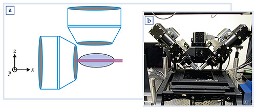
The light sheet itself can be generated in several ways. The two most common methods are to use a cylindrical lens to expand a laser beam in one dimension or to scan the pencil beam in a direction orthogonal to the propagation direction. The cylindrical-lens approach is easier to implement, but the resulting light sheet is not terribly uniform in intensity. Scanning a pencil beam generates a more uniform illumination; moreover, one can modulate the intensity to create a structured illumination that improves resolution. In addition, the scan can be synchronized with the operation of the detecting camera, a move that can further cut down on the out-of-focus light that is registered. On the other hand, with the scanning method, at any given instant, the camera records a line of light rather than a plane. So the scanning method is not as efficient as the cylindrical-lens approach. Also, scanning requires a more intense beam, and so it is the less gentle of the two principal approaches.
Two views are better than one
For wide-field, confocal, and light-sheet microscopy alike, the resolution detected in the axial direction is poorer than the resolution in the two other, orthogonal directions; that’s a worry for those of us trying to visualize submicron features in 3D. For example, in our work we follow the growth of individual neurons in C. elegans, and those cells often form a complex and twisted 3D structure within the embryo. We need good resolution in all dimensions.
To achieve that goal, we acquire two complementary views (the “dual view” of the diSPIM technique) by swapping the roles of the lens that produces the light sheet at the sample and the one that detects fluorescence for imaging onto the recording camera. A mathematical analysis (called joint convolution) of the combined data gives good resolution in all dimensions, as shown in figure 2a.

Figure 2. Dual-view inverted selective-plane illumination microscopy (diSPIM) in action. (a) In the left panel, beads are illuminated and recorded as in figure
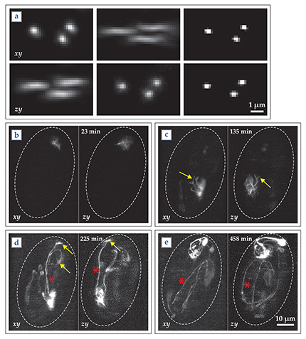
Our diSPIM approach has enabled us to clearly image the development of neurons in C. elegans throughout the entirety of embryogenesis. Figures 2b–2e show typical time-lapse images in which one can see structures as small as the “growth cones” that form as neurites develop. We are optimistic that eventually we will be able to use diSPIM to map the complete neurodevelopment of the embryonic C. elegans brain. Hopefully, the lessons learned from the roundworm and other organisms studied by our colleagues will apply to humans as well and thus help us better understand how our own brains form.
We thank members of the Shroff and Colón-Ramos research teams for helpful interactions and acknowledge with appreciation the assistance of the Institute of Neurobiology at the University of Puerto Rico; the Marine Biological Laboratory in Woods Hole, Massachusetts; and the Research Centers in Minority Institutions Program and the Intramural Research Program of the National Institutes of Health. The online version of this Quick Study includes an extended, annotated bibliography and a
References
1. Y. Wu et al., “Inverted selective plane illumination microscopy (iSPIM) enables coupled cell identity lineaging and neurodevelopmental imaging in Caenorhabditis elegans,” Proc. Natl. Acad. Sci. USA 108, 17708 (2011). https://doi.org/10.1073/pnas.1108494108
2. A. Kumar et al., “Dual-view plane illumination microscopy for rapid and spatially isotropic imaging,” Nat. Protocols 9, 2555 (2014). https://doi.org/10.1038/nprot.2014.172
3. WormGUIDES: Global Understanding in Dynamic Embryonic Systems, http://www.wormguides.org .
More about the authors
Abhishek Kumar is a postdoctoral fellow at Yale University in New Haven, Connecticut, and a guest researcher at the National Institute of Biomedical Imaging and Bioengineering (NIBIB) at the National Institutes of Health, in Bethesda, Maryland; Daniel A. Colón-Ramos is an associate professor of cell biology and cellular neuroscience at Yale; and Hari Shroff is chief of the section on high-resolution optical imaging and a tenure-track investigator at NIBIB.


