X rays from a free-electron laser resolve the structures of complex biomolecules
DOI: 10.1063/1.3580482
X-ray crystallography is remarkably successful at yielding atomic-resolution structures of proteins and other biological molecules. But that success has relied on growing macroscopic crystals. The countless identical molecules arrayed in a crystal share a radiation dose orders of magnitude higher than any one of them could tolerate alone. Moreover, the interference of x rays elastically scattered from the molecules concentrates the scattering intensity in a set of Bragg peaks. The larger the crystal, the better the signal-to-noise ratio.
Unfortunately, many biomolecules, particularly the proteins responsible for cross-membrane communication, resist forming macroscopic crystals; some defy crystallization altogether. In 2000, Uppsala University’s Janos Hajdu and colleagues simulated what would happen to a single uncrystallized protein or small assemblies of them placed in a hard x-ray beam from a free-electron laser (FEL). 1 According to their model, photoionization would destroy a protein in a few tens of femtoseconds. Stripped of electrons, the protein becomes highly charged and blows apart in what’s known as a Coulomb explosion. But if the x-ray pulses are sufficiently short and intense, they predicted, elastically scattered photons could yield useful structural information before the molecule or cluster starts to disintegrate. Paradoxically, the best way to image a pristine sample may be to destroy it.
In September 2009 SLAC’s Linac Coherent Light Source (LCLS) FEL began operation, packing nearly 1013 photons in each pulse at durations as short as 10 fs and peak power densities of 1016 W/cm2. Three months later, an international collaboration of more than 80 scientists led by teams from the German Electron Synchrotron (DESY), the Max Planck Institute for Medical Research, Arizona State University, and Uppsala used 1.8-keV x rays from the FEL for two proof-of-concept demonstrations of the idea.
The collaboration has just published the results: In one study, the research-ers resolved the structure of a photosynthetic membrane-protein complex (photosystem I) at a resolution of 8.5 Å, fine enough to identify its main structural features. 2 In another, performed the same week, they imaged a single virus—though at the far more modest resolution of 32 nm. 3 Both experiments represent a step toward making molecular movies of conformational dynamics with atom-by-atom detail using harder and shorter pulses.
One at a time
The achievements at SLAC build on work in 2006 that first tested Hajdu’s prediction and gauged the feasibility of running the experiment for real. Hajdu, Henry Chapman (both part of the SLAC collaboration), and colleagues used the FLASH facility at DESY, where Chapman now works, to deliver 25-fs laser pulses to a micron-sized picture etched on a silicon nitride membrane. Although the DESY experiment ran at a much longer wavelength in the extreme UV and at lower power than the new work, the single-shot diffraction pattern was rich enough to faithfully embody the picture’s structure before it was destroyed (see PHYSICS TODAY, January 2007, page 19 ).
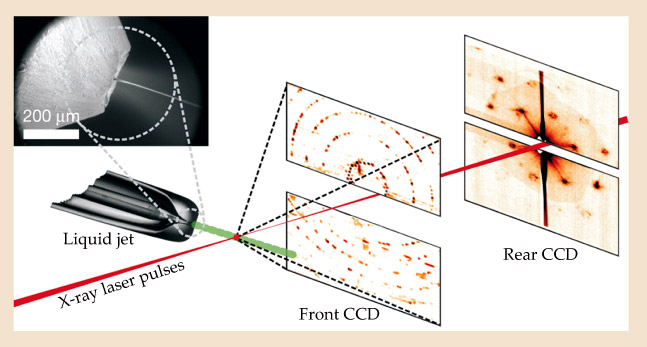
Unlike a solid membrane statically fixed in a beam, biomolecules are hard to pin down. To capture diffraction patterns in the new work, the collaboration devised a method to inject protein nanocrystals—and, separately, uncrystallized virus particles—into the FEL’s train of x-ray pulses. Figure 1 outlines the protein experiment. The nanocrystals, suspended in water to preserve their naturally hydrated structure, are squirted in a fine liquid stream across the beam. To prevent the stream from freezing in the vacuum chamber and to taper its width to match the beam’s, the researchers surrounded the stream with a coaxial flow of helium gas.

Figure 1. Serial nanocrystallography. A thin stream of protein nanocrystals held in liquid suspension is injected into vacuum and across SLAC’s free-electron laser beam. One at a time, the crystals intersect the pulsed x-ray beam at random times and orientations. Each hit vaporizes each crystal, but not before elastically scattered x rays escape; front and rear CCD detectors record the high- and low-angle diffraction peaks. Inset, a scanning electron micrograph shows the nozzle, liquid jet, and coaxial gas. (Adapted from ref.
The nanocrystals’ destruction hardly matters, as fresh ones are supplied after each pulse. Each time a nanocrystal intersects the beam, photons scattered from its reflection planes fill out the Bragg peaks. Over a few hours, hundreds of thousands of diffraction patterns are measured from randomly oriented crystallites of various shapes and sizes—each pattern a two-dimensional snapshot, or projection, of the protein complex in reciprocal space.
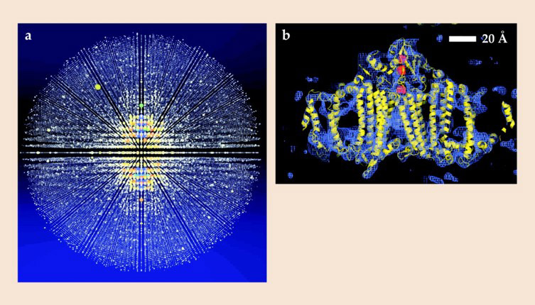
After deciphering the orientations of each sample using a crystallographic indexing process, the collaborators numerically merged 15 000 of the roughly 3 million collected snapshots into a high-resolution 3D diffraction pattern. The many contributions around each Bragg reflection from different nanocrystals add up to produce the Bragg peaks shown in figure 2.

Figure 2. (a) This projection of three-dimensional x-ray diffraction data comes from more than 15 000 single-crystal snapshots of the membrane-protein complex photosystem I, which comprises 36 proteins and 381 cofactors bound to them. (A movie that rotates the projection through the 3D volume is available with the online version of this article.) (b) Converting from reciprocal space to real space gives a 3D charge-density map. The experimental structure (blue) roughly matches the known best-estimate model (yellow and red) calculated from synchrotron x-ray crystallography of a macroscopic sample of photosystem I. Unlike nanocrystals, the macroscopic crystal had required more than 10 years of trial and error to synthesize. (Adapted from ref.
Fourier transforming the pattern into a real-space image requires not only the peaks’ intensities, which are captured by CCD detectors, but also the phases of scattered photons, which aren’t. Fortunately, those phases, which encode the locations of features, are not lost but reside in the fringes between Bragg peaks. They can be retrieved
using computer algorithms that iterate between real and reciprocal space.
In its work on photosystem I, though, the collaboration already knew the protein complex’s basic structure, derived from synchrotron data taken from a macroscopic crystal of the material years earlier. For its proof of concept, the researchers didn’t reconstruct the phases de novo, but simply computationally adjusted the positions of the protein complex’s known molecular structure until its calculated Bragg intensities reassuringly matched those they had recorded.
Single-shot imaging
At 0.75 µm wide, the mimivirus is the largest known virus, comparable in size to the smallest living cells. That’s too big for a full 3D reconstruction using cryo-electron microscopy (see the article by Robert Glaeser in PHYSICS TODAY, January 2008, page 48 ), and the fibrils surrounding the virus’s capsid prevent crystallization.
By using an FEL to image mimivirus particles, the collaboration could, as in the protein experiment, avoid staining, sectioning, freezing, or any other special preparations that might denature the structure. The experimental setup is also much the same, with one key difference: Viruses are injected across the x-ray beam as a focused jet of gas, rather than as a liquid stream, to reduce the background scattering.
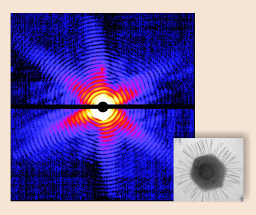
X-ray diffraction is incredibly inefficient. More than 99.9% of the photons don’t interact with a sample at all, and most of the others are annihilated when they eject photoelectrons. Even so, thanks to the FEL’s brightness—nine orders of magnitude greater than that of the best synchrotrons—nearly 2 million scattered photons contribute to each diffraction pattern, such as the one shown in figure 3. With that intensity, the team was able to reconstruct a 2D projection of the virus’s interior features—albeit at low spatial resolution—from a single laser pulse.

Figure 3. A single-shot diffraction pattern of a single mimivirus. The symmetry in the intensity pattern of concentric fringes mirrors that of the virus’s icosahedral capsid, evident in the inset electron micrograph of a thin section of the mimivirus. (Adapted from ref.
Unlike nanocrystals, whose orientations are straightforwardly determined from Bragg peaks, the diffraction pattern from a single object like a virus is continuous. At the time of the experiments, a low hit rate of mimivirus particles by the beam limited the quality of data the team could collect. With an improved injector and more than a million new diffraction patterns taken from SLAC in January of this year, the collaboration is now merging thousands of 2D patterns to reconstruct a full 3D image of the virus at higher spatial resolution.
The availability of the FEL as a tool for biologists has certainly tantalized them. Comments Stanford University’s Axel Brunger, “Its impact on structural biology could be similar to that of synchrotron light sources when first introduced for macromolecular-crystal data collection—a prerequisite for solving several prize-winning structures.”
References
1. R. Neutze, et al. Nature https://doi.org/NATUAS 406, 752 (2000).
2. H. N. Chapman, et al. Nature https://doi.org/NATUAS 470, 73 (2011).
3. M. M. Seibert, et al. Nature https://doi.org/NATUAS 470, 78 (2011).


