Two-Dimensional Optical Spectroscopy Identifies Crucial Intermolecular Couplings in Photosynthesis
DOI: 10.1063/1.2012448
The central step in photosynthesis, the transfer of energy to chemically active electrons, happens in a bulky protein complex called the reaction center. Feeding each reaction center with energy are hundreds of chlorophyll molecules that either convert sunlight into excitation energy or direct the energy, from molecule to molecule, toward the reaction center.
The chlorophylls, and the proteins that hold them, form light-harvesting systems that vary in shape and size across the span of photosynthetic life. But all the systems—in plants, algae, or photosynthesizing bacteria—share a remarkable ability: 99.5% of the solar energy they collect reaches their reaction centers.
Such efficiency is puzzling because the chlorophylls are crowded and coupled to each other. The chance of energy leaking out as phonons is conceivably high. But ironically, as Graham Fleming and his coworkers have now discovered, those couplings, rather than hinder efficient transfer, actually mediate it. 1
Fleming is the deputy director of the Lawrence Berkeley National Laboratory in California and holds a joint appointment at the University of California, Berkeley. He and his coworkers made their discovery using two-dimensional spectroscopy. Developed first for nuclear magnetic resonance (NMR) in the 1970s, the technique has only recently become practicable in the IR and optical. Indeed, Fleming’s group is the first to apply 2D optical spectroscopy to resolve coupled electronic transitions.
Fenna-Matthews-Olson
The photosynthesizing bacterium Chlorobium tepidum lives in hot smelly springs, where it uses red light to oxidize ambient sulfides. As light-harvesting systems go, C. tepidum’s is specialized and elaborate. However, one of the system’s key components is relatively simple and serves as a model system. Named after the scientists who isolated it and derived its structure, the Fenna-Matthews-Olson complex transmits electronic excitation energy to the reaction center via seven chlorophylls. Fleming’s collaborator Robert Blankenship of Arizona State University in Tempe isolated and purified the FMO used in the Berkeley study.
Excitation energy flows through FMO as electrons skip down the chlorophylls’ energy levels. The ease and rapidity of that flow underlies the high efficiency of photosynthesis. But identifying the actual paths taken by the electrons is difficult because the energy landscape of each chlorophyll is altered by FMO’s protein scaffold and by the other chlorophylls.
That landscape, as viewed with conventional 1D spectroscopy, consists of a string of broad peaks and troughs that neither resolve the individual levels nor reveal their mutual couplings. To get a sharper look, Fleming turned to 2D spectroscopy.
Two dimensions
Two-dimensional spectra map the signal transmitted by a sample as a function of two frequencies: excitation and detection. Roughly speaking, if the molecules in a sample don’t interact with each other, or if too much time elapses between excitation and detection, a 2D spectrum collapses to a 1D spectrum that lies along the diagonal. But when the molecules are caught interacting, distinct off-diagonal “cross peaks” appear.
Like the earliest and simplest 2D NMR schemes, the Berkeley experiment uses a sequence of three brief, broadband pulses. The first pulse excites all the transitions of interest. After what NMR spectroscopists call the coherence time τ, the sample is hit by a second pulse, which, depending on the value of τ and the details of the intermolecular coupling, either boosts or damps the signal at each resonant frequency. After a second interval, called the population time T, a third pulse probes the sample’s state, which decays over what’s called the detection time t.
The evolution of the intermolecular couplings is manifested in a sequence of 2D spectra extracted at different values of T. The axes of the spectra, τ and τ, are formed by Fourier-transforming the signal with respect to τ and t.
The signal whose analysis yields the spectra is not the third and final pulse, but a fourth, spontaneously created signal known as the photon echo. This signal, one of several manifestations of four-wave mixing, arises from the third-order nonlinear interaction of the three pulses with the oscillating molecular dipoles, the chlorophylls, in the sample.
Fourier transforming any signal requires keeping track of the phase. In the optical regime, where the field oscillates at frequencies of order 1015 Hz, doing so directly is impossible. Instead, one has to resort to interferometric techniques.
In a sense, the signals in 2D optical spectroscopy are already interferometric. As the first pulse passes through the sample, it induces the molecular dipoles to oscillate. The second pulse, if it’s not too late, will induce further oscillation that interferes with the first and creates what is, in effect, a diffraction grating for the third pulse. The photon echo emerges as the third pulse diffracts.
Extracting useful information from the photon echo depends crucially on controlling the phase of the three pulses throughout the experiment. At the radio frequencies used in NMR, phase is quite tractable. Translating the techniques of NMR to the optical regime seemed a distant, daunting feat. But in 1998, David Jonas of the University of Colorado in Boulder read a paper 2 by Theodor Hänsch and Ferenc Krausz and was inspired. Jonas realized that NMR-like control could be obtained simply by making sure that all three pulses have the same common envelope phase—that is, the same pattern of field oscillation. 3 Beam-splitting a single pulse achieves that.
Beam-splitting is also important for spatially isolating the weak photon echo from its three much brighter progenitors. Fortunately, the photon echo of three pulses that arrive from three different directions emerges in a predictable, momentum-conserving fourth direction. Although three carefully aligned beam splitters can create the desired pattern, Dwayne Miller of the University of Toronto in Canada showed last year that a micromachined diffractive optic will do the job more simply and with lower noise and greater stability. 4
The experiment
The Berkeley experiment was built and run by Tobias Brixner, Jens Stenger, and Igor Stiopkin. As their light source, they used their own, home-built Ti:sapphire laser, which produces femtosecond pulses centered on a wavelength of 550 nm.
At such short wavelengths, mitigating phase decoherence is essential. Miller’s diffractive optics scheme helps, as does the ingeniously simple method for introducing the two adjustable delays, τ and T. The team uses pairs of identical glass wedges. Pressed together, thin end to thick end, the wedges form a parallel-faced plate. Pulses pass through the plate’s center at right angles to the faces. Moving the wedges apart perpendicular to the pulse direction while keeping the faces parallel reduces the total thickness encountered by pulses and shortens the time delay.
To detect the photon echo, the team adapted a technique pioneered 10 years ago by Manuel Joffre of the Laboratory for Optics and Biosciences in Palaiseau, France. 5 The photon echo is combined interferometrically with a fourth pulse that traverses the sample in the same direction as the photon echo but ahead of it to avoid contributing to the echo.
The bottom panel in figure 1 shows the 1D spectrum (the dashed black line) along with the contributions (the dashed green lines) to the spectrum made by each of FMO’s seven excited states. Known as excitons, the excited states are collective quasiparticles. They provide a convenient and natural way to represent the energy landscape of a coupled system of molecules.

Figure 1. Two-dimensional spectra resolve coupled features that overlap in one dimension by spreading them out in a second. The bottom panel shows the 1D transmission spectrum of FMO (black) along with the seven excitons (green) that contribute to it. The top panels show 2D spectra at three different population times T. As T increases, the spectral contributions of the excitons, numbered 1 through 7, change.
(Adapted from ref. 1)
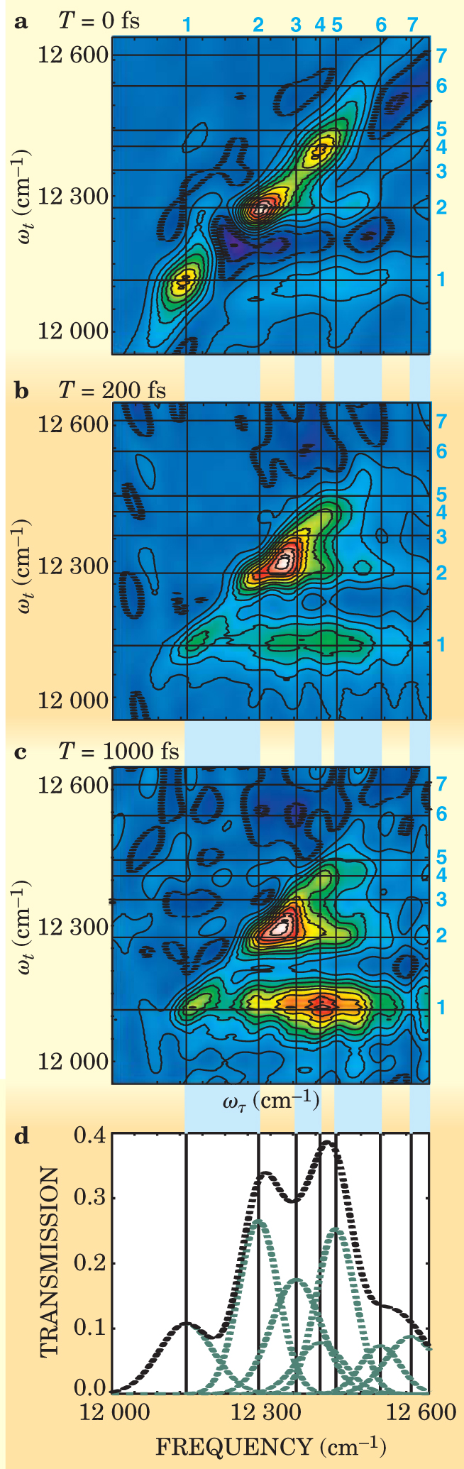
The top three panels in figure 1 show the 2D spectrum at three different values of T. Each spectral peak is formed by one or more excitons, whose central frequencies are indicated by the horizontal and vertical lines. When T = 0, that is, before energy flow begins, the peaks occupy the diagonal and more or less match the 1D spectrum in the bottom panel.
As T increases, to 200 fs in figure
Harsha Vaswami, Fleming’s newly graduated student, and Minhaeng Cho, his collaborator from Korea University in Seoul, simulated the experiment. Although their computer model missed some features, it does, in combination with the data, provide a consistent—and surprising—picture of the energy flow.
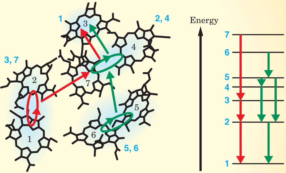
In general, the closer two states are in energy, the faster the transition between them. One might expect the electrons to hop from one level to the next lowest level and so on. But, as summarized in figure 2, that’s not what happens. In the two pathways the Berkeley team identified, electrons take advantage of the delocalized coupling between neighboring chlorophylls to skip between states that are close in space but not necessarily adjacent in energy. Doing so speeds the flow of energy, which is too fast for any energy-sapping phonons to respond.

Figure 2. FMO’s seven chlorophylls (numbered in black) are held in a certain configuration by FMO’s protein scaffold (not shown). As indicated by the pale blue shading, some of the chlorophylls’ excitons (numbered in blue as in figure
(Adapted from ref. 2.)
Phonons and other physical features limit the efficiency of photovoltaic cells to about 20%. Could the photosynthetic pathway be exploited to make a device that converts light into electricity with C. tepidum’s stunning efficiency? Copying its light-harvesting system wouldn’t work because the bacterium, like other photosynthesizing organisms, runs an elaborate biomolecular mechanism to mitigate and repair radiation damage. Conceivably, a bio—nano hybrid could be built. But, says Fleming, “it’s a distant dream.”
References
1. T. Brixner et al., Nature 434, 625 (2005) https://doi.org/10.1038/nature03429 .
2. L. Xu et al., Opt. Lett. 21, 2008 (1996) https://doi.org/10.1364/OL.21.002008 .
3. J. D. Hybl et al., Chem. Phys. Lett. 297, 307 (1998) https://doi.org/10.1016/S0009-2614(98)01140-3 .
4. M. L. Cowan, J. P. Ogilvie, R. J. D. Miller, Chem. Phys. Lett. 386, 184 (2004) https://doi.org/10.1016/j.cplett.2004.01.027 .
5. L. Lepetit, G. Chériaux, M. Joffre, J. Opt. Soc. Am. B 12, 2467 (1995) https://doi.org/10.1364/JOSAB.12.002467 .


