Nanodiamonds shine as subcellular thermometers
DOI: 10.1063/PT.3.4611

Once in a while when a diamond forms, a nitrogen atom steals the place of a carbon atom next to an empty carbon site in the crystal lattice. Researchers call such impurities nitrogen–vacancy (NV) centers and often deliberately add them to a diamond lattice for various applications, including as a component in quantum information technology or as a microscopic biological sensor. (See the article by Lilian Childress, Ronald Walsworth, and Mikhail Lukin, Physics Today, October 2014, page 38 .)
The sensing ability arises from the centers’ optical behavior. An NV center’s electronic ground state is a spin triplet, and the energy difference between the sublevel with spin quantum number 0 and the degenerate −1 and +1 sublevels is temperature dependent. The splitting can be measured using optically detected magnetic resonance spectroscopy. Shining a green laser on an NV center will raise one of its two localized electrons to the first excited state, and the NV center fluoresces red as the excited electron relaxes back to the ground state. Applying microwave radiation that is resonant with the sublevel splitting will cause the fluorescence intensity to decrease, and temperature can then be estimated by that difference in intensity.
The spin state of a nanodiamond NV center has been used as a magnetometer to detect weak magnetic fields in cancer cells (see Physics Today, August 2011, page 17 ). Nanodiamond NV centers have also been used as thermometers for in vitro cell cultures. 1 Now Masazumi Fujiwara at Osaka City University in Japan and his colleagues have demonstrated that NV centers can serve as precise quantum sensors to measure temperature changes in vivo in a complex organism. 2
By tracking nanodiamond particles with a microscope, the researchers monitored, with fine spatial and temporal resolution, the temperature inside the cells of Caenorhabditis elegans worms. Compared with quantum dots—another nanoscale temperature-taking tool and one that often contains cadmium and arsenic—nanodiamonds are less toxic to living organisms, and their chemical stability makes them less disruptive to various biochemical processes in cells.
Follow the light
For decades, scientists have used C. elegans in molecular biology research. The transparent, multicellular nematode resides in a sort of goldilocks zone of study: The animal is more complex than simple one-celled organisms, but not so complicated that researchers struggle to disentangle the effects and mechanisms of various biological machinery. Two Nobel Prizes in Physiology or Medicine were awarded for research on the nematode, for example—in 2002 for studies of organ development and cell death and in 2006 for RNA interference. What’s more, in December 1998 C. elegans became the first multicellular organism to have its genome sequenced.
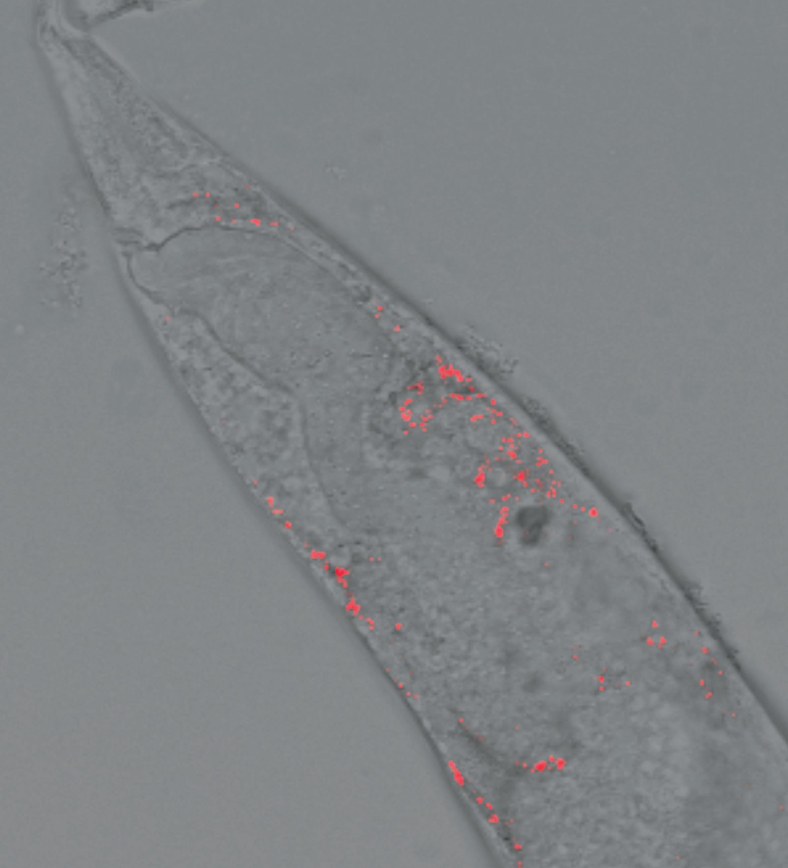
Fujiwara and his colleagues were interested in using the model organism to test an improved nanodiamond thermometer. They first injected nanodiamonds with NV centers into 1-mm-long C. elegans specimens, one of which is shown in figure
Figure 1.

Fluorescent nanodiamonds (red dots) were injected into a Caenorhabditis elegans worm about 80 µm in diameter. This image was captured with a confocal fluorescent microscope after the specimen was placed in a glass-bottom dish and the nanodiamonds were excited with a green laser. (Image courtesy of Masazumi Fujiwara.)
The nanodiamonds were excited by a green laser, and the fluorescence intensity of the NV centers was measured at four closely spaced microwave frequencies. That tactic provided the researchers with the means not just to estimate temperature but also to correct for errors associated with changes to the total fluorescence rate.
The researchers measured the fluorescence intensity along all three axes of the microscope every few seconds to track the NV centers through the worm body. Nanodiamonds don’t move all that much through in vitro cells. “Inside C. elegans or other animals, it’s a more dynamic environment,” says Fujiwara. “The technological breakthrough here was that we made a microscope system that can measure mobile nanodiamonds.”
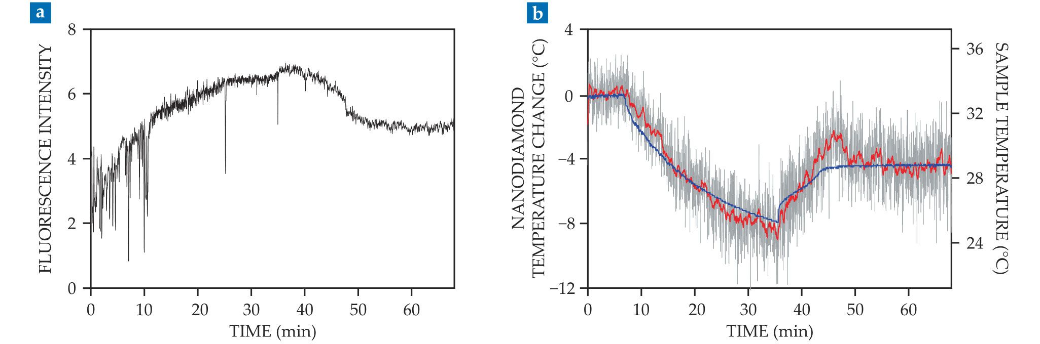
To test the sensitivity of their nanodiamond thermometer, the researchers subjected worms to thermal shocks, varying their body temperatures between 25 °C and 33 °C. Figure
Figure 2.

Monitoring subcellular temperature variations. (a) The fluorescence generated by the nitrogen–vacancy (NV) centers in a nanodiamond after excitation by a green laser. Temperature affects the transition energy between the sublevels of the NV center’s spin state and can be measured as an optically detected magnetic resonance when the excited electrons fluoresce as they relax back to the ground state. (b) The temperature estimated by the NV nanodiamond thermometer at 1 s intervals (gray line) and the 20-s running average (red line) are consistent with the independently labeled sample temperature (blue line). (Adapted from ref.
After observing induced temperature changes, the researchers used their nanodiamond thermometer to collect real-time measurements of heat generation with micrometer-scale spatial resolution. In a pharmacological experiment to treat cold exposure in a worm, the researchers introduced an uncoupling chemical that generates heat inside the mitochondria of a cell by interrupting the regular metabolic processes that operate there. Tracking the NV centers during the experiment showed that the worm moved a few micrometers, and the nanodiamond thermometer recorded a temperature increase of several degrees that persisted for about two hours.
Reconciling observations and theory
The temperature variations of a few degrees reported by Fujiwara and his colleagues agree with previous spectroscopic measurements of endogenous heat generation within a single living cell. But predictions based on the theoretical heat-generation rate and heat transfer in a cell showed temperature variations several orders of magnitude smaller. 3
“There’s no agreement among the researchers,” says Yutaka Shikano at Keio University in Tokyo, a coauthor of the new paper. “But we’ve measured the temperature robustly, and this is the temperature increase that we’ve found.” To close the gap between measured and predicted temperature changes, researchers will need to combine nanodiamond thermometry with other biological measurements, such as oxygen consumption rate. Such a combination of analyses promises to provide a better understanding of the biological mechanisms producing the temperature variations.
Besides C. elegans, Fujiwara and his coauthors suggest that a nanodiamond thermometer may also be useful for in vitro human stem cell studies. Researchers often use incubators to provide a lab environment for the cells to grow, but maintaining a stable temperature is challenging. Precise nanodiamond-based temperature measurements at the subcellular level could better determine local changes of the environmental temperature and help researchers better understand how those small differences affect cellular reproduction.
References
1. G. Kucsko et al., Nature 500, 54 (2013). https://doi.org/10.1038/nature12373
2. M. Fujiwara et al., Sci. Adv. 6, eaba9636 (2020). https://doi.org/10.1126/sciadv.aba9636
3. G. Baffou et al., Nat. Methods 11, 899 (2014). https://doi.org/10.1038/nmeth.3073
More about the authors
Alex Lopatka, alopatka@aip.org


