Lab Weds Brain Research and Physics
DOI: 10.1063/1.1955469

What are you thinking? Researchers at NeuroSpin won’t read your mind, but they do want to know how it works. The new facility near Paris will study the brain at previously untapped spatial and temporal scales.
Neurons can currently be probed a million at a time using magnetic resonance imaging (MRI), or by the dozen with electrical recordings. “We want to address the middle scale—clusters of 1000 to 10 000 neurons,” says founding director Denis Le Bihan, who holds a PhD in physics and a medical degree and is the driving force behind NeuroSpin. Going to higher MRI field strengths, he adds, will sharpen the resolution by about a factor of 10, from a few millimeters to hundreds of micrometers and from one second to hundreds of milliseconds.
Big biology
“The beauty of MRI,” Le Bihan says, “is that we have not reached the physical limits of the technique. That’s not true for other techniques, such as microscopy, where the resolution is given by the wavelength of light.” MRI is limited by the diffusion of molecules, he adds. “Diffusion will blur the images, but we are far from this.” Today, the strongest MRI machines for humans are 9.4 tesla—and only two are in use. Achieving 11.7 T, says Le Bihan, “pushes the technology to the limits, but we are not breaking new ground. This avoids taking too much risk.”
Still, it will be technically challenging, says Guy Aubert, former head of the French research agency CNRS who, now in Saclay with the French Atomic Energy Commission (CEA), is overseeing the building of a whole-body 11.7-T magnet for NeuroSpin. The lab will also have a 3-T scanner; a 17-T scanner for rodents; spectroscopy and microscopy tools; lab space for electronics, biology, neuro-physiology, and chemistry; and facilities for human clinical studies. Says Aubert, “I think it’s the first time—at least in France—that life science is building big equipment like in physics.”
Ground was broken for NeuroSpin in late January. The instruments are to be installed starting next year, and the facility should be fully functioning in 2009. Setting up NeuroSpin costs “several tens of millions of euros,” says Le Bihan, who will move his research from the Service Hospitalier Frédéric Joliot in Orsay. The CEA, the greater Paris region, and other public and private sources are footing the bill. “We are now knocking on the door of [the European Commission in] Brussels for money”—and to make NeuroSpin a European user facility, Le Bihan says.
The facility will house some 160 researchers, half permanent and half visitors. Says Le Bihan, “Chemists will develop contrast agents and tracers. We need physicists, mathematicians, computer scientists—new software will be developed to analyze the huge amount of data that will be produced—neuroscientists, electro-physiologists, psychiatrists. Physicists and engineers from CEA will provide specific knowledge on magnetism, cryogenics, radio frequencies, and management of large projects. Theoretical physicists might be attracted because there is a need to build a working model of the brain.”
From magnets to mental illness
Research planned for NeuroSpin ranges from magnet development to cognitive studies to mental illness, brain development, and brain dysfunction. The high magnetic fields may shed light on autism, language development, aging, how the mind produces and appreciates art, and early detection of neurological disorders such as Alzheimer’s and Parkinson’s diseases. “It’s like getting a microscope to look at the brain noninvasively in three dimensions,” says Le Bihan.
For example, by using transgenic mice, “we hope to see how genes and the environment interact during brain development,” says Le Bihan. “Many brain disorders in adults can be the consequence of what happens during early stages of brain development, including [during] pregnancy.”
In gray matter, Le Bihan says, “when neurons get activated, they change shape. They increase their volume a tiny bit, and the water mobility decreases. If you can monitor this change in diffusion, you may be able to determine or infer which regions of the brain are being activated.” Diffusion MRI also promises to help unravel disorders affecting white matter—the fibers that connect different regions of the brain—such as dyslexia, multiple sclerosis, and schizophrenia.
Another approach, MRI and spectroscopy of heavier nuclei such as sodium, phosphorus, carbon, or oxygen—instead of hydrogen, the MRI workhorse—opens the way for broader study of brain metabolism, neurotransmission, and disease. “We use carbon-13 and oxygen-17,” says Le Bihan. “The abundance of those nuclei is very low, hence the need for very high fields.”
“I think [NeuroSpin] is a wonderful facility,” says Kamil Ugurbil, director of the Center for Magnetic Resonance Research at the University of Minnesota. “They are trying to combine and facilitate interactions between many different types of scientists. Such efforts go on already, but it’s based on individual efforts. By consciously planning this, both in terms of building design and in terms of organization, they may be able to achieve much better results.”

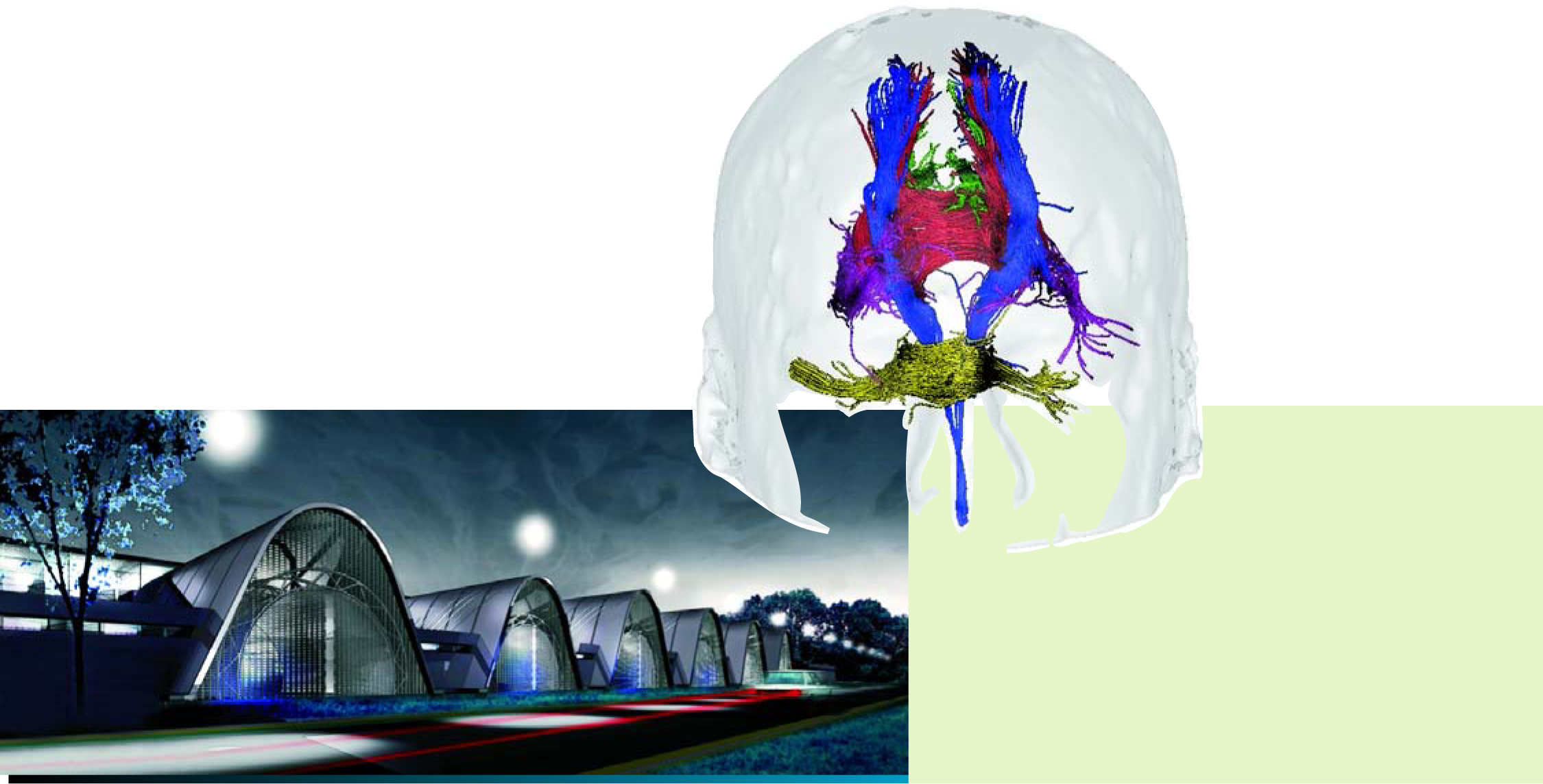
Modeling the brain will be the focus at NeuroSpin (left), an interdisciplinary lab being built in Saint Aubin, 40 km southwest of Paris. The facility’s strong magnetic fields will, for example, yield finer detail in in-vivo visualizations of the brain’s white matter (shown in false color, above) with diffusion magnetic resonance imaging.
BRAINVISA/UNAF/CEA/(Rendering by architect Claude Vasconi, reproduced courtesy of CEA.)

Le Bihan
N.SOUIRY
More about the authors
Toni Feder, American Center for Physics, One Physics Ellipse, College Park, Maryland 20740-3842, US . tfeder@aip.org


