Biological tissue can behave like a liquid crystal
DOI: 10.1063/PT.3.3580
Consisting of a small number of cell layers, an epithelium is the protective membrane that lines animal organs and embryos. The epithelium must continually renew itself, and its cells form a dense colony whose local tension fluctuates during morphogenesis, growth, and tissue repair as some cells are born and others die. Despite the ubiquity of those processes, however, the mechanical and biochemical mechanisms that regulate tensional homeostasis and determine which cells are targeted for removal have long been obscure. Dying or injured cells are not the only ones extruded. As biologists discovered in 2012, so are healthy cells whose migration or proliferation overcrowds the tissue. 1
Intercellular signaling, adhesive junctions, and dynamic stresses generated by the motion of cells are all thought to influence the targeting. But their interplay has made it difficult to disentangle relative contributions. Researchers from the Mechanobiology Institute (MBI) at the National University of Singapore, the Jacques Monod Institute at CNRS and Paris Diderot University, the University of Oxford, and the Curie Institute have now discovered that the arrangement of cells in the membrane may be the primary factor behind the mechanics and location of cell removal. 2
The discovery builds on earlier efforts in soft-matter physics to model biological tissue as a liquid crystal. Like collagen-producing fibroblasts, smooth muscle, lipids, and other biological cells, certain types of epithelial cells are rod shaped—longer than they are wide. When isolated, the cells move randomly but persistently, powered by their internal hydrolysis of adenosine triphosphate. But in dense colonies, the anisotropic shape prompts them to align into domains, much like rod-shaped nematic liquid crystals, whose orientational order lowers their collective free energy. Also like liquid crystals, the cellular packing contains defects—misalignments in the long-range orientation—that punctuate the separate domains.
When the MBI researchers and their colleagues cultured epithelial cells from a canine kidney into monolayer sheets and imaged them under a microscope, they were not surprised to find pervasive misalignments. The surprise was the defects’ profound influence on cell behavior. An analysis of the microscopic snapshots of the cultures revealed that the vast majority of cells extruded from the tissue came from lattice sites that hosted a comet-shaped defect—a misalignment in which cells in the “head” lie perpendicular to those in the “tail.” According to Benoit Ladoux, one of the principal investigators in the collaboration, the implication was immediately clear: “The positions of those defects represent a powerful predictor of where cells are likely to die.”
The usual suspects
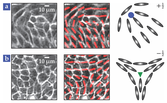
Orientational defects in a collection of motile cells emerge spontaneously as the cells flow en masse. Created in pairs, the defects can migrate apart and annihilate each other on recombining. Epithelia host comet-shaped and triradius varieties, shown in figure

Figure 1. Misalignments between rows of elongated cells in epithelial tissue take on the same structure as topological defects in nematic (orientationally ordered) liquid crystals. The top and bottom sequences show microscope images of two types of defects: (a) a comet-shaped (+½) defect, in which a row of cells in the “head” is oriented perpendicular to a row in the “tail,” and (b) a triradius (−½) defect, in which different rows diverge from each other. The cells’ calculated orientations (red, middle) reveal the resemblance to liquid-crystal defects (right), whose cores (blue and green) represent points of maximum misalignment. (Adapted from ref.
In some cases, the defects can self-organize into an ordered configuration. 4 In others, they can channel a one-dimensional flow of cells between themselves. This past month Harvard Medical School’s Kyogo Kawaguchi and his colleagues at the University of Tokyo and Kyoto University found that ±½ defects control the collective flow of neural stem cells cultured on a glass slide: The neural cells become depleted in the vicinity of a triradius and flow toward a comet-shaped defect. 5 An apparent logjam at the head of the comet causes cells flowing from the tail to accumulate into ever-denser 3D mounds. According to Kawaguchi, the channeled flow covers several millimeters and mimics the migration of cells in part of an adult mammal’s brain.
Defects kill
Although epithelial cells are packed more densely than neural stem cells and are more subtly rod shaped, they still migrate toward comet-shaped defects at about the same velocity—tens of microns per hour. To look for a causal role in the correlation found between defect sites and cell-extrusion events, Ladoux and his colleagues scanned relevant areas of the epithelial sheet using traction force microscopy. In that technique, fluorescent particles are embedded in a substrate under the sheet. The forces generated by the cells are determined from the measured displacement of the particles. Having measured those forces, the researchers were able to infer the compressive stress acting on cells near a defect.
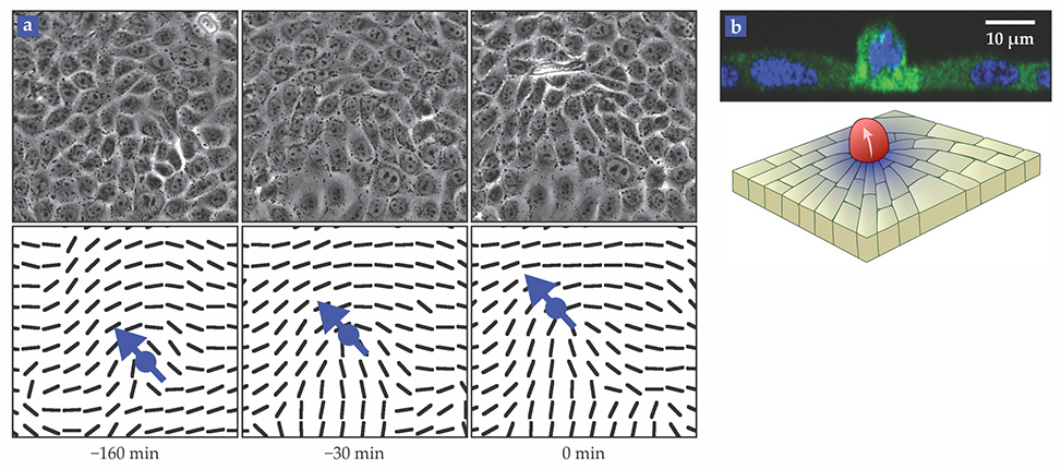
The measurements and simulations made by the researchers offered a consistent picture: The distortion on a cell caused by a misalignment with neighbors builds up for hours prior to an extrusion event, as cells in the tail of the comet overcrowd those in the head. And if the compression is high enough, it triggers an extrusion. The phase-contrast images shown in figure

Figure 2. Time evolution of a comet-shaped defect. (a) The top panels show a series of microscope images that capture dynamical changes to epithelial cells of a canine kidney in the same 100 µm by 100 µm scene. Bottom panels represent the calculated orientations (black lines) of the cells, modeled as components of a liquid-crystal lattice. The core (blue dot) of the defect migrates upward in the direction of the arrow. Cells in the comet tail become compressed against cells in the head. At a modest threshold stress of about 70 Pa, a cell located in front of the defect core is extruded (white). (A movie is available in the online version of this report.) (b) A side-view fluorescence image of an extruding cell is shown above a schematic of the scene. (Adapted from ref.
Further corroboration for that extrusion mechanism came from monitoring the activation of YAP, a stress-triggered protein in the cells’ cytoplasm, that the researchers observed in the vicinity of a defect. They also observed greater-than-normal levels of caspase 3, an enzyme that signals a cell’s impending death.
A complex choreography commences between a dying cell and its neighbors. A contractile cable forms around the cell and acts as a purse string to draw in surrounding cells to gently pinch off the dying piece. The cable is made of the scaffolding protein actin and the motor protein myosin; their coordinated motion provides the force behind the pinching.
The relative force contributions from actin–myosin and the defect compression that triggers cell death are not clear. Compression squeezes the cell, but that alone is unlikely to launch it outward like a tiddlywinks disk. The compression also activates enzymes that degrade cellular proteins, fragment the nucleus, and weaken the cell’s adhesive bonds with its neighbors. Those processes lead to the detachment of the dying cell from the tissue. As the purse string tightens, the neighboring cells stretch to prevent a gap.
Intriguingly, a phenomenon akin to extrusion also happens at defects in a generic liquid crystal. Any elongated object, whether an inanimate particle or a biological cell, becomes geometrically frustrated at the core of a topological defect (the blue and green shapes in figure
Defect control
Although defects emerge spontaneously in biological tissue, it’s possible to control their density—and thus the density of extrusion hot spots. Canine kidney cells prefer to align tangentially to the boundary of a substrate, and the researchers cultured their cells in one experiment atop a star-shaped substrate, as shown in figure

Figure 3. Geometry-induced defect production. (a) A star-shaped monolayer of cells produces comet-shaped topological defects predominantly at the tips of its four arms. (b) With their higher density of defects, those regions extrude a greater number of cells (red). The color denotes the normalized extrusion number. (Adapted from ref.
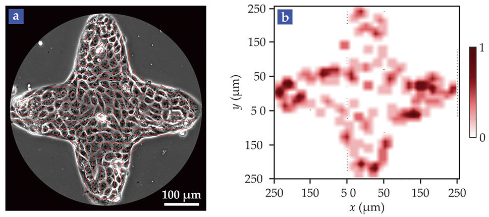
By contrast, the triradius defect density was higher near the center of the star, but without a commensurate increase in extrusion events there. The experiment further corroborates the causal link: The emergence of comet defects provides hot spots of compressive stress, which lead to a higher probability of cell death.
The demonstrated control over defect density may chart a path for scientists trying to suppress the invasion of cancerous tumors and other pathological tissue in which the process of cell death has gone awry. To influence the process, one can imagine inserting implants surgically to guide cell orientations in a way that produces topological defects.
Could such an approach work? “Why not?” quipped biophysicist Guillaume Charras at the London Centre for Nanotechnology. “I wonder about its practicality as a therapy, though. The usual problem is detection. And if you can detect a cancerous lesion, the most effective strategy is often to just cut it out with a scalpel.”
References
1. E. Marinari et al., Nature 484, 542 (2012);https://doi.org/10.1038/nature10984
G. T. Eisenhoffer et al., Nature 484, 546 (2012). https://doi.org/10.1038/nature109992. T. B. Saw et al., Nature 544, 212 (2017). https://doi.org/10.1038/nature21718
3. R. Kemkemer et al., Euro. Phys. J. E 1, 215 (2000);https://doi.org/10.1007/s101890050024
for a more recent account of defects in biological cells, see G. Duclos et al., Nat. Phys. 13, 58 (2017). https://doi.org/10.1038/nphys38764. S. J. DeCamp et al., Nat. Mater. 14, 1110 (2015). https://doi.org/10.1038/nmat4387
5. K. Kawaguchi, R. Kageyama, M. Sano, Nature (in press), doi:https://doi.org/10.1038/nature22321 .
6. M. Kleman, O. D. Lavrentovich, Soft Matter Physics: An Introduction, Springer (2003).


