Medical imaging with antimatter
DOI: 10.1063/pt.pswd.fhis

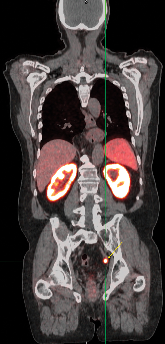
Most diseases manifest with biochemical signatures well in advance of anatomical changes. The image below shows a hybrid positron emission tomography/computed tomography (PET/CT) scan of a prostate cancer patient. The average-sized lymph node in the pelvis indicated by the yellow arrow would look normal in an anatomical image from a CT or magnetic resonance imaging (MRI) scan. But the PET scan shows, definitively, that the lymph node has been invaded by cancer cells: Before the scan, the patient was injected with a positron-emitting radiopharmaceutical that binds to an antigen found on the membranes of prostate cells, both normal and cancerous. PET imaging with disease-specific radiopharmaceuticals is dramatically changing the management of cancer through its remarkable sensitivity in detecting even tiny, remote tumors.

(Image courtesy of the University of Iowa department of radiology.)
The ability to image biochemical and physiological processes in real time distinguishes PET from medical imaging technologies that primarily visualize anatomy. 1 PET imaging can reveal the molecular signatures of a vast array of diseases and help physicians devise appropriate therapeutic strategies. Over the past four decades, PET has gradually evolved into a mainstream clinical diagnostic tool for cancer and cardiac and neurological diseases. More than 2 million scans are performed in the US annually, and approximately double that are performed worldwide.
A PET scan of glucose metabolism in the body of a breast cancer patient is illustrated in figure
Parts of the body that metabolize glucose appear darker, in proportion to the level of that metabolism. The scan shows normal physiological glucose metabolism in the brain, liver, and heart; intense localization in the urinary bladder and kidneys is because the kidneys recognize the glucose analog as an impostor and gradually filter it into the urine (a similar effect can be seen in the PET/CT scan of the prostate cancer patient). Most importantly, the scan identifies the presence of rapidly dividing metastatic tumor cells that avidly metabolize glucose in lymph nodes around the left armpit.
Basics of positron decay
PET gets its name from the nuclear radioactive decay mechanism—positron emission—that enables the localized measurement of biochemical activity in the body. A positron is an antimatter electron: It has the same mass as an electron but is positively charged. It most commonly comes from the radioactive decay of a proton-rich nucleus, in which a proton is converted into a neutron, as shown in figure
In that annihilation interaction, the electron and positron disappear. In their stead, two 511 keV gamma-ray photons, traveling at 180° from each other, are emitted from the point of annihilation. It is an elegant real-world example of Einstein’s E = mc2: The masses of the electron and positron (9.11 × 10⁻³¹ kg each) are converted into energy in the form of photons. The photons travel in opposite directions as a result of the conservation of momentum because both the electron and positron are functionally at rest when they interact and annihilate. PET scanners are designed to simultaneously—within about five-billionths of a second—detect both photons to determine the position of the decay event in the body.
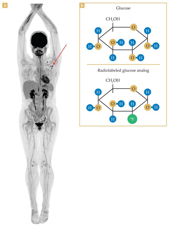
Figure 1.

Oncology using positron emission tomography. (a) A PET image of glucose (sugar) metabolism in the body of a subject with breast cancer is achieved by injecting a glucose analog radiolabeled with fluorine-18. Parts of the body that metabolize glucose appear darker. The image shows normal glucose metabolism in the brain, liver, and heart; the intense but normal localization in the urinary bladder and kidneys are because the kidneys recognized the analog as an impostor and gradually filtered it into the urine. Crucially, the scan identifies rapidly dividing metastatic tumor cells that avidly metabolize glucose in lymph nodes around the left armpit (red arrow). (b) The structure of a glucose molecule and the subtly altered structure incorporating a positron-emitting 18F atom. (Image courtesy of the EXPLORER Molecular Imaging Center/University of California, Davis.)
Radiopharmaceuticals
The key to PET imaging’s remarkable versatility is grounded in its molecular approach. In PET imaging, relatively short-lived radionuclides that decay by positron emission are chemically incorporated into molecules of physiological interest and introduced into the body, usually through an injection. The molecules, commonly called radiopharmaceuticals, could be neuroreceptor ligands designed to bind to dopamine receptors in the brain, 3 or they could be small molecules engineered to attach to specific proteins on the cell membrane of a particular cancer type. Or they could be as simple as radiolabeled glucose molecules used to image sugar metabolism in the body.
To be used in PET imaging, the positron-emitting radionuclide attached to the molecule needs to have a half-life long enough to allow for several steps of production and use: the drug’s radiochemical synthesis, subsequent quality control to ensure its safety and purity, its administration to a patient, and its sufficient circulation through the body to reach its molecular target and be imaged. Ideally, the half-life is tens of minutes to several hours. But sometimes, the physiological process being imaged requires days, in which case a longer-lived positron-emitting radionuclide, like zirconium-89 with a half-life of 78 hours, is necessary.
PET is a tracer-imaging methodology. Although it involves injecting drugs into a patient, the actual amount of the radioactive drug is incredibly small, far below the threshold of what would induce any kind of measurable pharmacological effect. For example, the mass of a standard clinical dose of the 18F-labeled glucose used for a PET oncology scan is about one-billionth of a single grain of sugar. That is enough to generate an excellent clinical-quality image, with more than 100 million pairs of annihilation photons detected in a typical 15-minute scan. The use of trace quantities of such drugs allows for the probing of biochemical distributions in the body noninvasively without perturbing the biochemical mechanisms that are being studied.
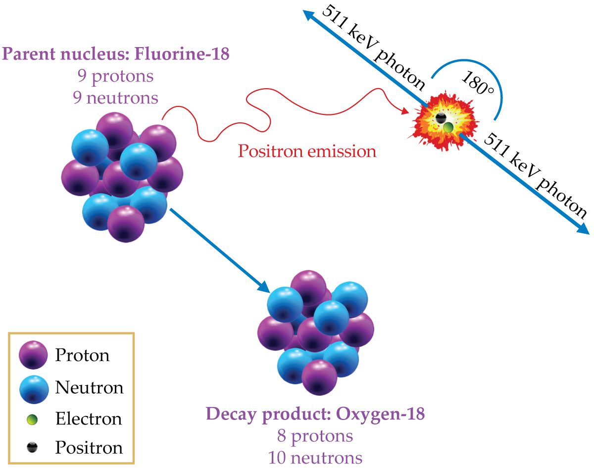
Figure 2.

Positrons are emitted in the decay of fluorine-18 to oxygen-18 when a proton in the 18F nucleus gets converted to a neutron. Within less than a millimeter of its original location, the positron will annihilate with an ambient electron. That annihilation produces two 511 keV gamma-ray photons, traveling in opposite directions, that can be detected by scintillation crystals in the ring of a PET scanner. Tens of millions of photon detections are compiled to generate PET scan images.
Approximately 90% of PET scans are performed to help diagnose and stage various kinds of cancer and to help monitor patients’ responses to treatment. The remaining 10% of clinical cases are split evenly into cardiac and neurological applications. The technology is also used extensively in drug development with specially designed ultrahigh-resolution PET scanners for small animals, especially rats and mice.
PET, CT, x-ray, and single-photon nuclear medicine imaging all use ionizing radiation to generate diagnostic images that help physicians manage their patients’ treatment. The amount of radiation energy deposited in organs and tissues with each of those procedures is well known and understood, and each procedure and protocol is optimized to provide diagnostic-quality imaging with the lowest associated radiation dose. For all those diagnostic procedures, the radiation doses received by a patient are orders of magnitude below any that could potentially cause a short-term adverse radiation effect. The primary concern about low radiation doses in diagnostic procedures is an exceedingly low increased risk of the patient developing a downstream cancer years later. It is a basic tenet of medical imaging that the benefit of the tests far outweighs any remote potential risk.
How a PET scanner works
A PET scanner is designed to generate a 3D map of the radiopharmaceutical’s distribution in the body. To achieve that, the scanner needs to identify the location of each positron decay event in 3D space with as much precision and efficiency as possible. The higher the precision with which the origin of the decay can be determined, the better the resolution can be of the resulting image. And with a higher detection efficiency, less of the radioactive drug needs to be injected to create a diagnostically satisfactory image.
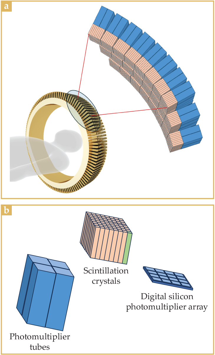
Figure 3.

A PET detector ring. (a) A zoomed-in section of the detector ring shows that it is made of many detector modules. (b) A traditional detector module is made of approximately 64 scintillation crystals (one here highlighted in green) that are optically coupled to photomultiplier tubes (PMTs). When a gamma-ray photon triggers a burst of scintillation light in a crystal, the relative amounts of light detected by each of the four PMTs will determine which crystal was involved in the event. New scanners couple the same scintillation crystal block matrix to a digital silicon photomultiplier array instead of to PMTs. Because fewer crystals are coupled to each photodetector, crystal identification is more reliable.
Because the emitted positron travels only a short distance (less than a millimeter for 18F) before annihilation, detecting the positron itself is out of the question. The two gamma-ray photons produced by the positron–electron annihilation, however, are highly penetrating. A reasonable fraction of the photons escape the body, unscathed by either Compton scattering or photoelectric-effect absorption. It is the efficient simultaneous detection of those photon pairs that is at the heart of the PET scanner’s design.
A modern-day PET scanner consists of multiple contiguous detector rings that contain thousands of tiny scintillation crystals, typically about 4 mm × 4 mm on the face and 15–25 mm in depth, as shown in figure
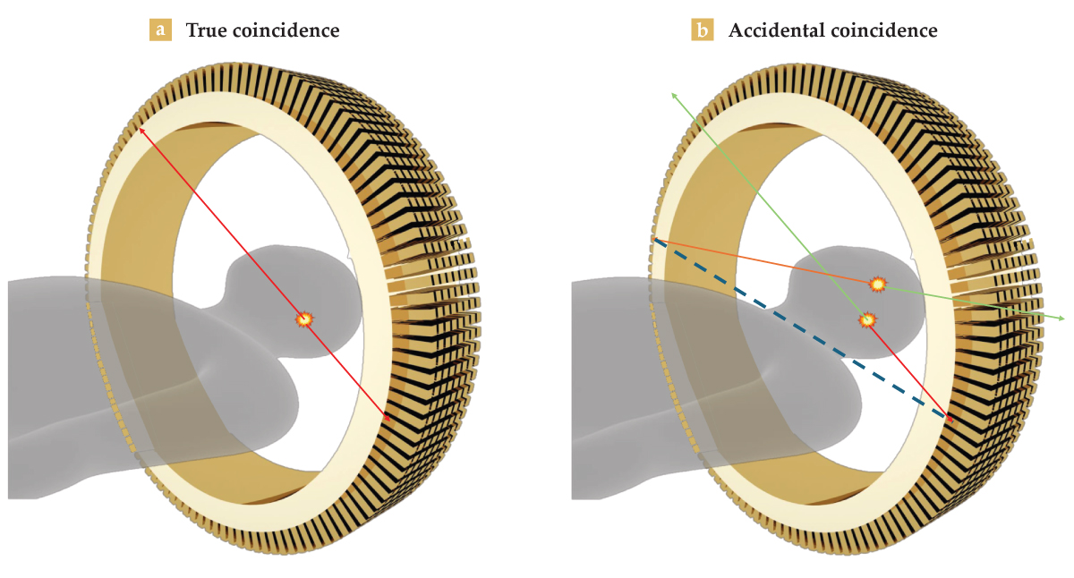
Following the detection of any single 511 keV photon, the PET system patiently waits about 5 ns for a second such photon. If a pair of 511 keV photons are detected within that 5 ns window, then the system concludes that the photons must have come from the same annihilation event, one that happened somewhere along a straight line between the two detectors, as illustrated in figure
Coincidences are rejected if one of the photons has an energy of less than 511 keV, which signals that the lower-energy photon was scattered. If two 511 keV photons are detected but not within the 5 ns coincidence window, they are rejected because they must have originated from different decay events. It is possible that two independent decays occur within 5 ns of each other and that unrelated 511 keV photons are simultaneously detected, which creates an accidental or random coincidence. The random coincidence, if left uncorrected, would create the illusion that a decay event occurred along the dotted line in figure
A deeper dive into detection
There are three main components to the detector modules used in PET: the scintillation crystal that converts the gamma-ray energy to visible light, the photodetector that converts the visible light to an electrical signal, and the detector electronics that measure the absorbed energy and record the precise time and position of the detection event. The three components need to work together to detect the origin of the radioactive decay with high spatial resolution, high efficiency, high energy resolution, and high temporal resolution.
As shown in figure
Detector modules are configured in multiple contiguous rings to allow simultaneous collection of imaging data along the length of the patient. Most PET scanners can image between 20 and 25 cm along the axis of the scanner at a time, though designs are moving toward the inclusion of more rings and therefore a larger axial coverage beyond 25 cm. The patient is slowly moved through the scanner to image as much of their body as is medically necessary.
To precisely identify the location from which the positron–electron annihilation event originates, PET requires simultaneous gamma-ray detection with high spatial resolution. High resolution is primarily achieved by decreasing the size of the face of the individual scintillation crystals. Modern PET systems typically have square crystal faces with sides measuring 3 to 4 mm and a depth of about 15 to 25 mm. With crystals that size, clinical resolution can approach 4 to 5 mm. The longer the crystals are, the higher the probability that an incoming 511 keV photon interacts with them. But longer crystals have a higher risk of the photon crossing over from one crystal into an adjacent crystal before interacting, which will result in a small misplacement of where the annihilation event occurred and a slight decrease in resolution.
Figure 4.

Photon coincidence detection in a PET scanner. (a) When two 511 keV gamma-ray photons are detected within 5 ns of each other, they are presumed to have formed from the annihilation of a positron and an electron somewhere along a straight line between the two detection locations. That measurement corresponds to a true coincidence event. (b) It is possible for two discrete annihilation events to randomly occur within 5 ns of one another such that two unrelated 511 keV photons are detected at the same time. Those accidental coincidence events are indistinguishable from true events, but they can be approximated statistically and subtracted at the end of a scan.
The more efficient the radiation detectors are in a PET scanner, the less radioactive material needs to be injected into the patient to still achieve a diagnostic-quality image. High detection efficiency is achieved by using scintillation crystals with a high average number of protons in the nucleus and high physical density. Detection efficiency is further enhanced by simply using thicker crystals. A PET crystal with a depth of 20 mm has about an 80% probability of detecting a 511 keV gamma ray.
When a scintillation crystal is hit with a single high-energy photon, it can fully absorb that energy through the photoelectric effect and convert the energy into several thousand visible-light photons that produce a rapid flash inside the crystal. The number of generated visible-light photons is proportional to the energy absorbed, so by measuring the amount of light produced, one can indirectly measure the energy of the incoming photon. That capability is critical because photons that experience inelastic Compton scattering on the way to the detector will have lower energy and can thus be filtered out.
PET image reconstruction
PET images are created through image-reconstruction algorithms that use the tens of millions of 511 keV photon pairs detected during the scan. At the foundation of PET reconstruction is the concept of back projection. If an annihilation event is sensed between two detectors, the position of the annihilation event is assigned an equal probability over the entire length of the line connecting them. That back projection is done for each detected event. Where events’ individual back projections spatially overlap, their probabilities are summed. When all detected events are collected and overlapped, it results in a first approximation of the radioactivity distribution.
The smaller the detectors and the finer the angular sampling, the higher the resolution of the resulting image. And although coincidence windows of about 5 ns are typically used, modern PET detection systems can achieve timing resolution on the order of 200–300 ps. Light travels only about 6 cm in 200 ps. By measuring the difference in arrival times between two coincident photons, the position of the annihilation event can be reasonably localized to within several centimeters along the line connecting the two detectors. The refined localization based on that time-of-flight information greatly enhances the probability-based reconstruction algorithm so that substantially better images are produced from fewer detected events. 4
Back projection is only the first step of what is a much more complex reconstruction process. The entirety of the collected projection data is corrected for photon attenuation within the body, electronic dead time, accidental coincidences, and scattering. Finally, just before the back-projection step, the projection data from each angle—that is, all the parallel lines connecting opposite detectors at a given angle—are processed through a spatial filter that largely removes the artificial blurring introduced in the back-projection step. That processing step is also used for other tomographic imaging methods, including CT scans. (See reference for an excellent animated demonstration of the back-projection method.) Reconstruction steps are typically performed iteratively to converge on the highest-quality clinical images.
When all those steps are performed on a calibrated PET system, the resulting image is a quantitative map of radioactivity concentration, which reveals the spatial distribution of the radiopharmaceutical in the body. Each voxel, or 3D pixel, in the image is in units of becquerels per cubic centimeter, where the becquerel is one radioactive decay per second. In well-calibrated systems, measurements can be accurate to better than 2%. Beyond being remarkably useful medical imaging tools, PET scanners are amazing measurement devices, and for decades, they have provided accurate pharmacokinetic measurements of numerous physiological processes.
Necessary infrastructure
The clinical use of PET scanners has experienced continuous growth over the past 30–35 years; millions of diagnostic scans are currently being performed annually. That number, however, is one to two orders of magnitude lower than the number of annual CT and MRI scans being performed. Growth of PET usage has been more gradual than for its CT and MRI counterparts largely because of infrastructural and regulatory barriers.
PET radiopharmaceuticals are considered drugs by the US Food and Drug Administration and other worldwide regulatory agencies. As such, before clinical use, a PET radiopharmaceutical must undergo testing in phase 1, 2, and 3 clinical trials to prove both safety and clinical efficacy. Clinical trials cost tens of millions of dollars to perform, and approval of a radiopharmaceutical for human clinical use can easily take 5–10 years.
Additionally, PET drugs are radiolabeled with relatively short-lived radionuclides that have half-lives typically on the order of an hour or two, which means that they must be synthesized near to both the time and place they are used. A PET-dedicated cyclotron is typically necessary to produce the positron-emitting radionuclides that are incorporated into the PET radiopharmaceuticals. For PET to be widely available requires a network of commercial radiopharmacies equipped with cyclotrons, radiochemistry laboratories, clean-room facilities for drug manufacturing and quality control, and centralized distribution infrastructure.
It has taken some time to build that infrastructure. At present, there are hundreds of PET-dedicated cyclotrons around the world, daily producing carbon-11 (a half-life of about 20 minutes), 18F (a half-life of about 110 minutes), copper-64 (a half-life of about 13 hours), and 89Zr (a half-life of about 78 hours) to supply the thousands of worldwide PET scanners.
The cutting edge
PET imaging continues to grow and evolve. It has become increasingly useful in clinical medicine. One area of advancement is through technical innovation—making PET scanners with higher resolution, more sensitivity, and enhanced image quality through better time-of-flight resolution and use of AI in the image-reconstruction phase.
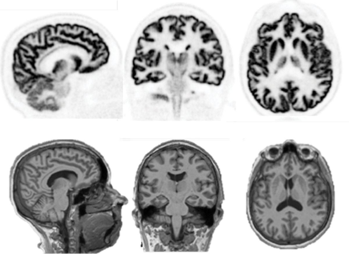
Figure 5.

Image slices of the brain of a healthy control participant (top row), acquired 60–90 minutes after injection of a radiolabeled synaptic density marker. The images were made using a new dedicated human-brain PET imager, NeuroEXPLORER. Matching MRI anatomical images were also captured (bottom row). The ultrahigh resolution and sensitivity of the NeuroEXPLORER provide exquisite delineation of the folds of the brain’s cerebral cortex and structures deeper in the brain. (Image courtesy of Richard Carson on behalf of the NeuroEXPLORER consortium.)
One trend in new commercial equipment is to have more detector rings to produce a longer axial field of view. With the additional rings, oncology patients can be scanned faster and with improved image quality. Or the image quality and scan time can be kept the same and smaller radiopharmaceutical doses used, which is especially important for reducing radiation exposure in pediatric patients. Some commercial PET systems on the market have axial fields of view of 1–2 meters that can image a patient’s entire body in just a few minutes; the scan in figure
Meanwhile, developments in electronics and photosensor technologies for silicon photomultipliers continue to improve time-of-flight resolution, which now is better than 200 ps and provides enhanced image quality. 7
New brain-only imaging units are being developed with an unprecedented isotropic resolution that approaches 2 mm3, which is comparable to but not quite as high as that of MRI. PET brain images, like those shown in figure
The true power of PET imaging lies in its ability to probe the specific physiology of disease processes. In the clinical domain, only about 20 PET radiopharmaceuticals are approved for clinical use. But those well-studied and proven drugs only scratch the surface; hundreds of PET radiopharmaceuticals under development are aimed at new molecular targets. Drugs under later-stage development include a class that specifically binds to fibroblast activation protein, a marker found on cells in the tumor microenvironment of a broad array of cancers.
Another area of growth is the use of PET to image and monitor the body’s immune response. 8 There are several strategies to do that, but one of the more direct ways is to label and monitor monoclonal antibodies, which are synthetic proteins similar to the body’s natural antibodies. Monoclonal antibodies are used to treat cancer because they bind to specific antigens on cells and trigger immune responses. Because they are typically large molecules that have slow kinetics, they sometimes take days to localize, so longer-lived positron emitters, such as 89Zr, are often used to allow for imaging several days after injection.
Neurological and psychiatric radiopharmaceuticals are also clinically available, with more under development. With the recent approvals of new treatments for Alzheimer’s disease, demand for PET imaging to more definitively identify candidates for therapy is rising dramatically. 9 On the research and drug-development front, radiopharmaceuticals designed to measure the pharmacokinetics and distribution of positron-emitter-labeled molecules targeting dopamine and serotonin receptors are popular targets for study. And direct measurement of neuroinflammation with PET has applications in studying the biophysiology and biochemistry of stroke, Parkinson’s disease, and traumatic brain injury, among other disorders.
Molecular imaging with PET is poised for continued rapid growth, driven by advancements in imaging technology, an increasing array of disease-specific radiopharmaceuticals, and its alignment with the worldwide focus on personalized medicine.
This article was originally published online on 14 August 2025.
References
1. M. E. Phelps, J. Nucl. Med. 41, 661 (2000) https://jnm.snmjournals.org/content/41/4/661 .
2. J. S. Fowler, T. Ido, Semin. Nucl. Med. 32, 6 (2002).https://doi.org/10.1053/snuc.2002.29270
3. N. D. Volkow et al., J. Nucl. Med. 37, 1242 (1996) https://jnm.snmjournals.org/content/37/7/1242.long .
4. J. S. Karp et al., J. Nucl. Med. 49, 462 (2008).https://doi.org/10.2967/jnumed.107.044834
5. A. Kesner, “3D image reconstruction,” Human Health Campus, International Atomic Energy Agency (2016), https://humanhealth.iaea.org/HHW/MedicalPhysics/NuclearMedicine/ImageAnalysis/3Dimagereconstruction/ .
6. R. D. Badawi et al., J. Nucl. Med. 60, 299 (2019).https://doi.org/10.2967/jnumed.119.226498
7. P. Lecoq, S. Gundacker, Eur. Phys. J. Plus 136, 292 (2021).https://doi.org/10.1140/epjp/s13360-021-01183-8
8. W. Wei et al., Chem. Rev. 120, 3787 (2020).https://doi.org/10.1021/acs.chemrev.9b00738
9. P. N. E. Young et al., Alzheimer’s Res. Ther. 12, 49 (2020).https://doi.org/10.1186/s13195-020-00612-7
More about the authors
John Sunderland is a medical physicist and a professor of radiology and physics at the University of Iowa in Iowa City. He is also the director of the university’s PET Imaging Center.


