Surface chemistry experiments speed up
DOI: 10.1063/PT.3.2134
In studying a chemical reaction on a surface, it’s useful to know not only the identities of the desorbed products but also their three-dimensional velocity distributions. The distribution of kinetic energies provides information about the energy released in the reaction. And the details of any anisotropy in the product velocities offer clues into the transition-state geometry and forces imparted to the products.
Ion-imaging techniques developed for gas-phase chemistry, which capture products on a 2D position-sensitive detector, can help to measure velocity distributions. But until recently, limitations on imaging speeds have meant that only one product could be detected at a time. Repeating the experiment to look for every possible product—and a reaction involving organic molecules can have many—is tedious. And for a surface reaction, it requires the preparation of new, clean surfaces, which can complicate comparisons of data from different repetitions.
Over the past several years, a team of chemists and physicists at Oxford University and the Rutherford Appleton Laboratory (RAL) developed a new ultrafast sensor—called PImMS, for pixel imaging mass spectrometry—that allows the imaging of multiple products in a single experiment. 1 So far, it’s been used mostly for gas-phase chemistry. Now Michael White and colleagues, at Brookhaven National Laboratory and Stony Brook University, have applied PImMS to surface chemistry. 2 And the technique shows promise for many more applications.
Imaging ions
The basic concepts of ion imaging date back decades. 3 A chemical reaction is initiated, typically by a pulsed laser, and the products are ionized and accelerated by an electric field toward the detector. Microchannel plates convert the incident ions into bursts of electrons, and a phosphor converts the electrons into an optical signal, which can be captured by a camera, typically a CCD. In 1997 André Eppink and David Parker modified the technique by refining the ion optics, a series of charged electrodes that focus all product molecules with the same initial velocity vector onto the same spot on the detector, even if they originate in different places. 4 That velocity-map imaging technique, which allowed the reaction to be carried out in a larger region without loss of image resolution, proved extraordinarily useful to the field of gas-phase chemical dynamics.
Acceleration in an electric field separates the ionized products by their mass-to-charge ratio, and almost all the ions are singly charged, so different chemical species arrive at the detector at different times; in that respect, the technique resembles time-of-flight mass spectrometry. But the different products are separated by at most a few microseconds, whereas a commercial CCD has a time resolution of tens of milliseconds. By time-gating the instrument’s electronics, one can use a CCD to capture an image of a single product mass, but not to separately image more than one mass in a single cycle.
Enter PImMS
Oxford chemists Claire Vallance and Mark Brouard were experienced in both ion imaging and mass spectrometry, and the PImMS collaboration arose from their desire to combine the two. They teamed up with Oxford physicist Andrei Nomerotski (and later Richard Nickerson), who brought expertise in detectors for particle physics, and Renato Turchetta of RAL, a specialist in sensor design.
Unlike a CCD, the PImMS camera doesn’t capture a series of full-frame images. At the desired 25-ns frame rate, most of those images would be blank. Instead, each time one of the camera’s 5184 pixels detects a flash of light, it records a time-stamped data point, (x, y, t). The electronics that produce the time stamps—more than 600 transistors per pixel—are contained in the pixels themselves. 5 Such complex circuitry needs to be shielded from parasitic collection of the charge carriers generated by the incoming photons. That shielding is achieved by strategically placing implants of highly doped semiconductor, a technology known as INMAPS (isolated n-well monolithic active pixel sensors) from Turchetta’s group. 6 INMAPS is also being deployed in particle-physics detectors.
Vallance and Brouard did the first test experiments of PImMS on gas-phase systems. Last fall, Nomerotski went to Brookhaven to work on the detectors for the Large Synoptic Survey Telescope. There he met with White, who expressed an interest in the PImMS camera. White had long known about the impact that ion imaging had had on the study of gas-phase reactions, and he wanted to try it out on surface reactions. He’d made some preliminary attempts using a conventional camera, but with limited success. The PImMS camera, which allowed imaging of all product masses simultaneously, was just the thing he’d been looking for.
Surface chemistry
For their proof-of-principle experiment, White and his group looked at photooxidation of 2-butanone (CH3COCH2CH3) on titanium dioxide, a material known for its ability to catalyze the degradation of organic pollutants into water, carbon dioxide, and smaller organic molecules. Previous studies of that reaction had identified strong signals at 27, 28, and 29 atomic mass units, corresponding to the organic fragments C2H3, C2H4, and C2H5. But it wasn’t clear whether each of those fragments was really produced in the surface reaction or whether some of them might be the result of larger fragments falling apart when ionized. Such dissociative ionization is a pervasive phenomenon that complicates the study of almost all organic photochemical reactions.
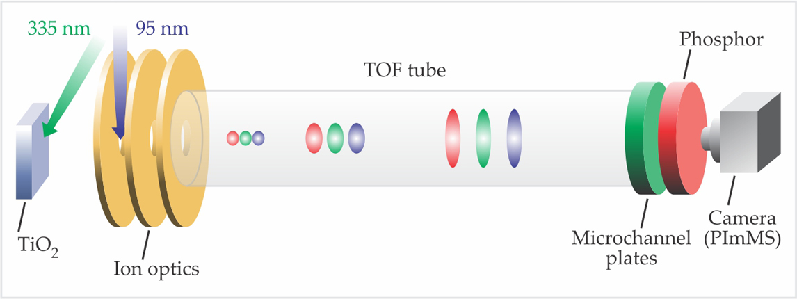
White and colleagues prepared a single-crystal surface of TiO2, dosed it with O2 and 2-butanone, and irradiated it with pulsed 335-nm laser light to initiate the reaction. With their ion optics set up for velocity-map imaging, they could irradiate the whole centimeter-square surface and still obtain angular information on the desorbed products. The products were ionized with 95-nm radiation, as shown in figure 1, and accelerated toward the detector and the PImMS camera.

Figure 1. Instruments for imaging a surface reaction, shown schematically. Pulsed laser light at 335 nm initiates a reaction among molecules adsorbed on the titanium dioxide surface. Desorbed products are ionized by 95-nm radiation, focused by the ion optics, and accelerated toward the detector. As they traverse the time-of-flight (TOF) tube, products spread out in space and become separated by their mass-to-charge ratio. Each ion incident on the microchannel plates creates a burst of electrons. The phosphor converts the electron burst into an optical signal, which is imaged by the camera. (Adapted from refs.
Some of their data are shown in figure 2, with the detector’s two spatial dimensions collapsed into one for ease of display. Signals appear, as expected, at masses 27, 28, and 29, with the lower-mass signals appearing larger on the detector plane. That spatial size progression was entirely consistent with dissociative ionization: C2H5 radicals are produced in the surface reaction, and some of them, when ionized, lose one or two of their hydrogen atoms along with an electron. With the momentum imparted to them by the escaping H atoms, the C2H3 and C2H4 fragments end up with broader transverse velocity distributions than their parent C2H5 radicals.

Figure 2. Pixel imaging mass spectrometry data on the photooxidation of 2-butanone on a titanium dioxide surface. The two dimensions of the detector plane are collapsed into one for ease of viewing. (Adapted from ref.
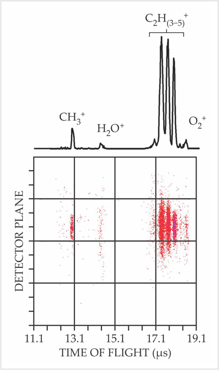
But the smoking gun was in the C2H3 signal. The spatial distribution of data at mass 27 is so broad, compared to the finite size of the ionizing region, that it could not result from C2H3 radicals desorbed directly from the surface. It must be due to dissociative ionization.
Moving ahead
White and colleagues plan to continue to use PImMS to study surface photochemistry. The camera’s time resolution and multimass capabilities will allow the study of velocity and spatial product distributions in complex polyatomic systems. The researchers’ long-term goal is to combine PImMS with ultrafast lasers to watch surface reactions in real time.
Photochemistry—in the gas phase or on a surface—is not the only possible use for the PImMS camera. As Vallance and Brouard emphasize, it can be used for any particle-imaging application that requires nanosecond to microsecond time resolution. For example, by refocusing the ion optics so that each pixel collects all particles with a common point of origin rather than a common velocity vector, one can obtain mass-resolved images of surfaces, including biological specimens. 7
References
1. A. T. Clark et al., J. Phys. Chem. A 116, 10897 (2012). https://doi.org/10.1021/jp309860t
2. M. D. Kershis et al., J. Chem. Phys. 139, 084202 (2013). https://doi.org/10.1063/1.4818997
3. D. W. Chandler, P. L. Houston, J. Chem. Phys. 87, 1445 (1987). https://doi.org/10.1063/1.453276
4. A. T. J. B. Eppink, D. H. Parker, Rev. Sci. Instrum. 68, 3477 (1997). https://doi.org/10.1063/1.1148310
5. J. J. John et al., J. Instru. 7, C08001 (2012).
6. J. A. Ballin et al., Sensors 8, 5336 (2008). https://doi.org/10.3390/s8095336
7. M. Brouard et al., Rev. Sci. Instrum. 83, 114101 (2012); see also http://pimms.chem.ox.ac.uk . https://doi.org/10.1063/1.4766938
More about the authors
Johanna L. Miller, jmiller@aip.org


