Physics meets biology at new HIV structural biology centers
DOI: 10.1063/1.2883903
Angela Gronenborn is a trained physicist, with a specialty in nuclear magnetic resonance spectroscopy. But when her biologist brother started working on cellular proteins, she decided she’d put her NMR skills to work helping to figure out the structures of protein–DNA complexes. “That’s when people said I was crazy,” she states.
Nowadays, Gronenborn is the principal investigator at the University of Pittsburgh Center for HIV Protein Interactions, one of three HIV structural biology centers that were established in August 2007 with $54 million in grants from the National Institutes of Health (NIH). Fully half of the researchers who are affiliated with the center were trained as chemists or physicists, she says. The major tools the center is using in the quest to understand the mechanisms by which HIV infects human host cells and hijacks them to replicate itself originated from the physical sciences. By elucidating the molecular structure of the viral proteins and the ways they interact with proteins in the human immune-system cells they infect, the center researchers expect to locate targets for drug designers to use in their quest to render HIV harmless.
The Pittsburgh center and those at the University of Utah and the University of California, San Francisco (UCSF) are jointly funded by five-year grants from NIH’s National Institute of General Medical Sciences and National Institute of Allergy and Infectious Diseases (NIAID). Ravi Basavappa, a structural biologist and program director for the biophysics branch at NIGMS, says physics is central to studying the dynamics of the interactions that occur between HIV and human proteins. He estimates that 70% of the scientists affiliated with the centers would describe themselves as biophysicists, in that they apply biophysical techniques in their research.
Blocking HIV replication
Although the centers are working on separate pieces of the protein puzzle, their collective goal is to find methods to prevent HIV proteins from functioning effectively. They are trying to determine the structures of the host-cell protein complexes that govern HIV replication, in the hope that they can block those interactions, explains Diana Finzi, program officer in the AIDS division at NIAID. The most straightforward approach targets the proteins produced by the virus themselves. The current generation of drugs embody this approach by blocking the functions of enzymes known as reverse transcriptase and protease.
But with a cure for AIDS still elusive, researchers have moved on to the more difficult approach of inhibiting the functions of complexes that comprise two or more proteins. The focus of the centers is to determine the three-dimensional structures of those complexes and to define the areas in their complicated molecular structures where they interact and therefore are vulnerable and can be interfered with. The task is complicated by the constant mutations HIV undergoes and by the fact that the virus thrives in an activated immune system, Finzi says. The trick will be finding a target that will block HIV proteins without harming the cell, she adds.
Indispensible tools in the centers’ work are x-ray crystallography, NMR spectroscopy, and cryo-electron microscopy. Advancing the state of the art for those imaging technologies, and making them freely available to the scientific community, are expected to be a spinoff benefit.
X-ray crystallography, the workhorse of structural biology, employs x-ray diffraction to determine the molecular structure of proteins that have been crystallized. A half-dozen synchrotron radiation sources at national laboratories and universities are the brightest sources of experimental x rays and a good deal of crystallography is done there.
Limits of crystallography
Growing crystals is both time-consuming and technically challenging, and many proteins, notably the roughly 30% of known proteins that reside in cell membranes, aren’t water soluble and can’t be crystallized. Enter NMR spectroscopy, which doesn’t require crystals and offers an additional capability to see proteins move in response to changes in their environment. NMR also can be used to determine which amino acid residues are involved in protein–protein interactions, without scientists’ having to solve the structure of the entire protein complex.
But NMR is currently limited to defining the structures of proteins that contain about 300 or fewer amino acids—structures about 60 times smaller than the largest molecules that have been resolved by crystallographers. NMR also is expensive and difficult to adapt to high-throughput methods, says Basavappa, and requires longer data collection times,
Gronenborn, a well-known NMR spectroscopist, acknowledges that the technology is less mature than x-ray crystallography, which has evolved to the point where it is considered “black box,” meaning experimenters don’t need to be conversant in the technology to use it.
A third technique
A third technique, cryo-electron microscopy, produces a 3D image from multiple images of a frozen sample that are taken from varying angles. (See Physics Today, January 2008, page 48 .) While the other two techniques provide an averaged picture of assemblies, cryo-EM is capable of assessing structural homogeneity. But its nanometer-resolution capability is lower than the angstrom-level resolution of the other two technologies and has generally limited cryo-EM use to the study of multiple-protein complexes. A few researchers are attempting to push cryo-EM to the point of resolving the amino acid building blocks of a protein, says theoretical chemist and biophysicist Greg Voth, a member of the Utah center. Voth and Alan Frankel, UCSF’s principal investigator, each say that improvements to the technology will be especially crucial to his center’s goals.
Single-molecule imaging
David Millar, a biophysicist at the Scripps Research Institute who is part of the Utah team, is using single-molecule fluorescence imaging to decipher the mechanism by which HIV’s genetic material is transported from the host cell’s nucleus back into the cytoplasm, where the cell’s protein factories are hijacked to produce more of the virus. By color-labeling each of the numerous proteins involved in that transport, Millar’s lab has been able to observe the sequence of events involved in formation of the large protein complex that transports the viral RNA within the cell.
Millar says the fluorescence technology will be useful for studying many other biological processes, including the mechanism by which ribosomes, the protein factories of the cell, are themselves assembled from some 20 individual proteins and RNA.
The centers are also using multiscale computer simulation, isothermal titration calorimetry, surface plasmon resonance, and fluorescent resonance energy transfer.
The interdisciplinary character of the centers allows the quantitative perspective of the physical scientists to be brought to bear on the biologists, who are more apt to express their findings in descriptive terms, says Gronenborn. “It’s very difficult for biologists to put their observations into quantitative terms.”
An arranged marriage
“We’re bringing together two very different worlds to apply what they know to the problem,” says Finzi. The arranged marriage is not without challenges, particularly regarding communication. “We speak different languages, and we don’t always understand what the others are doing,” says Finzi, who confesses that she has only a basic grasp of the imaging techniques.
The key to interdisciplinary efforts, Voth says, is to focus on one’s own area of expertise and let other collaborators do the same. Voth admits that he’s not aware of a single “card-carrying physicist” at the Utah-based center.
Frankel says three of the nine principal scientists in his collaboration, including an NMR spectroscopist and a computational expert, have strong physical science backgrounds. Most of the rest are biochemists.
But for all the expertise that’s being drawn into the centers, there’s no guarantee of success, Finzi cautions. “We think the centers have tremendous potential,” she says. “But it’s a little like gambling; sometimes you get something marginal, and sometimes you get a big hit.”

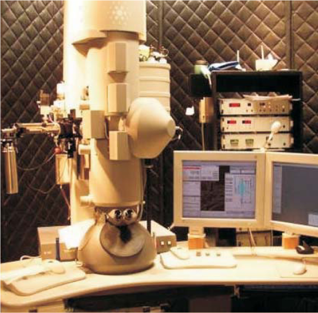
Cryo-electron microscopes are used to determine the structures of protein complexes that are key to HIV replication in human host cells.
WESLEY SUNDQUIST/UNIVERSITY OF UTAH
More about the authors
David Kramer, dkramer@aip.org


