Doubt is cast on a mechanism of cancer nanomedicine
DOI: 10.1063/PT.3.4426
Cancer is a disease of tissue growth gone wrong. Tumor cells proliferate uncontrollably and don’t die as they should. When a solid tumor grows large enough to need to sprout its own blood vessels, those vessels, too, grow irregularly. The tumor vessel networks are tortuous and disorganized (see Physics Today, February 2016, page 14 ), and the vessels’ lining, or endothelium, is riddled with gaps hundreds of microns wide between cells.
The challenge of chemotherapy is to kill off the tumor cells without doing too much harm to healthy ones. The inter-endothelial gaps promised a way to do that. Nanoparticles up to 300 nm in diameter can fit through the gaps, and they can’t permeate normal blood vessels the same way. So nanoparticles loaded with a drug should, it stands to reason, selectively enter and attack tumor tissue but leave healthy tissue alone. An inherently biomedical challenge was seemingly transformed into one of nanotechnology and fluid dynamics.
A quarter century of nanomedicine research has indeed yielded a few nanoparticle drug formulations that offer better performance or fewer side effects than their molecular counterparts. (See the article by Jennifer Grossman and Scott McNeil, Physics Today, August 2012, page 38 .) The mechanism of their action, however, has never been fully verified. No technique exists that can image nanoparticles in vivo, in real time, and with sufficient resolution to observe the particles slipping through the inter-endothelial gaps in a tumor’s blood vessels. The idea that they do has always been an assumption.
New research by Shrey Sindhwani, Abdullah Muhammad Syed (both with Warren Chan’s laboratory at the University of Toronto), and their colleagues suggests that that assumption might not be correct. Most nanoparticles that end up in tumors, the researchers found, get there by passing through the endothelial cells, not between them. 1
The underlying biological details are still murky. Several possible mechanisms fall under the umbrella of trans-endothelial pathways, and the researchers don’t yet know which one predominates or why. For example, individual endothelial cells might engulf the nanoparticles and then expel them into the surrounding tumor, or the nanoparticles might pass through small intracellular pores that open up to exchange nutrients across the endothelium.
What the possible mechanisms have in common, however, is that they’re all active cellular processes, so they’re sensitive to nanoparticle surface chemistry and composition in ways that a passive physical mechanism isn’t. The task of getting nanomedicines into tumors may therefore be a biological one rather than a physical one after all.
Mind the gaps
Chan and his colleagues have been testing the foundations of cancer nanomedicine for several years. In a controversial 2016 analysis of the literature, they found that among all published experiments in the field, a median of just 0.7% of the injected nanoparticles were delivered to the tumor. 2 Critics countered that some nanomaterials are delivered in quantities much greater than the median, that even 0.7% of the injected dose might be enough to treat a tumor, and that the amount of nanomaterial detectable in a tumor isn’t necessarily the best measure of a drug’s effectiveness. 3 But as Sindhwani explains, the Toronto researchers had their minds on a more fundamental question: “If there are all these gaps in the blood-vessel lining, why is the tumor accumulation so low? What are these gaps, and how many are there?”
Meanwhile, Sindhwani was also learning about Harold Dvorak’s work on trans-endothelial mechanisms that can carry proteins as large as 5 nm into tumors. “That work is relatively unknown in the nano community,” he says, “and nobody had looked at its relevance to nanoparticles.”
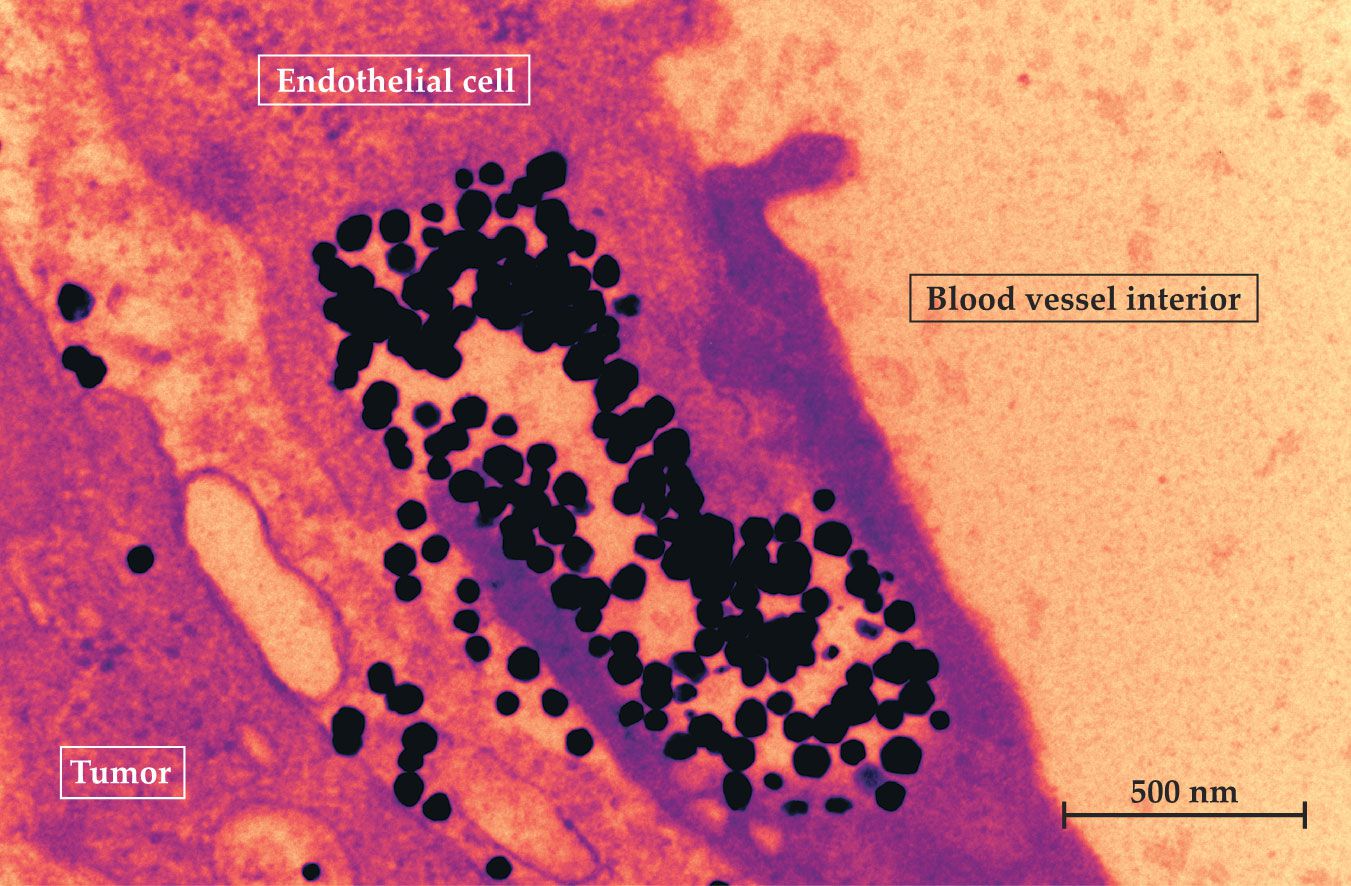
It’s relatively straightforward to show that trans-endothelial delivery of nanoparticles to tumors is possible. The researchers injected gold nanoparticles into a tumor-bearing mouse, allowed them to circulate, then extracted slices of the tumor and imaged them with transmission electron microscopy (TEM). Some of the images, including the one in figure
Figure 1.

Gold nanoparticles (black) injected into the blood of a tumor-bearing mouse appear in the cytoplasm of the endothelial cells that line the tumor blood vessel; a few have been expelled into the tumor. Nanoparticles have been assumed to enter tumors through large gaps in the endothelium, but no such gap is visible in this transmission electron microscopy image. (Courtesy of Shrey Sindhwani.)
But which of the possible mechanisms predominates? That’s a statistical question, and answering it required a lot of data—and a combination of methods. “It is really challenging to design experiments for looking at what nanoparticles are doing in the tumor,” says Syed.
The first step was to determine how many inter-endothelial gaps there really were. The researchers took TEM images of hundreds of blood vessels from both mouse and human tumors, and they trained a team of nine colleagues to identify the inter-endothelial gaps. To reduce bias, they had each image independently evaluated by three randomly chosen team members who didn’t talk to one another.
As it turned out, inter-endothelial gaps were surprisingly rare: Images of 313 blood vessels from mice showed just seven gaps in total, or about one per 20 000 square microns of endothelium. Human tumor vessels showed gaps in similarly small numbers. In a fluid-dynamical simulation of 50 nm nanoparticles, the observed density of gaps could account for just 1–2% of the already small number of nanoparticles known to reach the tumors.
Zombie mice
The researchers also wanted an experiment that could distinguish passive from active nanoparticle transport. For that, they developed a mouse model they called Zombie. Tumor-bearing Zombie mice, as the name suggests, are effectively animated corpses: dead, chemically treated to halt all cell activity, and hooked up to an external pump to keep their blood circulating. If nanoparticles enter tumors by passively flowing through inter-endothelial gaps, then just as many nanoparticles should reach the tumors in Zombie mice as in control, or live, tumor-bearing mice.
But that’s not what happened. Fluorescence images in figure
Figure 2.

Fluorescence images show the propagation of nanoparticles in the tumors of control tumor-bearing mice, which were alive, and Zombie mice, all of whose cells were dead. In the control mouse, nanoparticles (green) are able to escape the blood vessels (red), but in the Zombie mouse they can’t. (Adapted from ref.
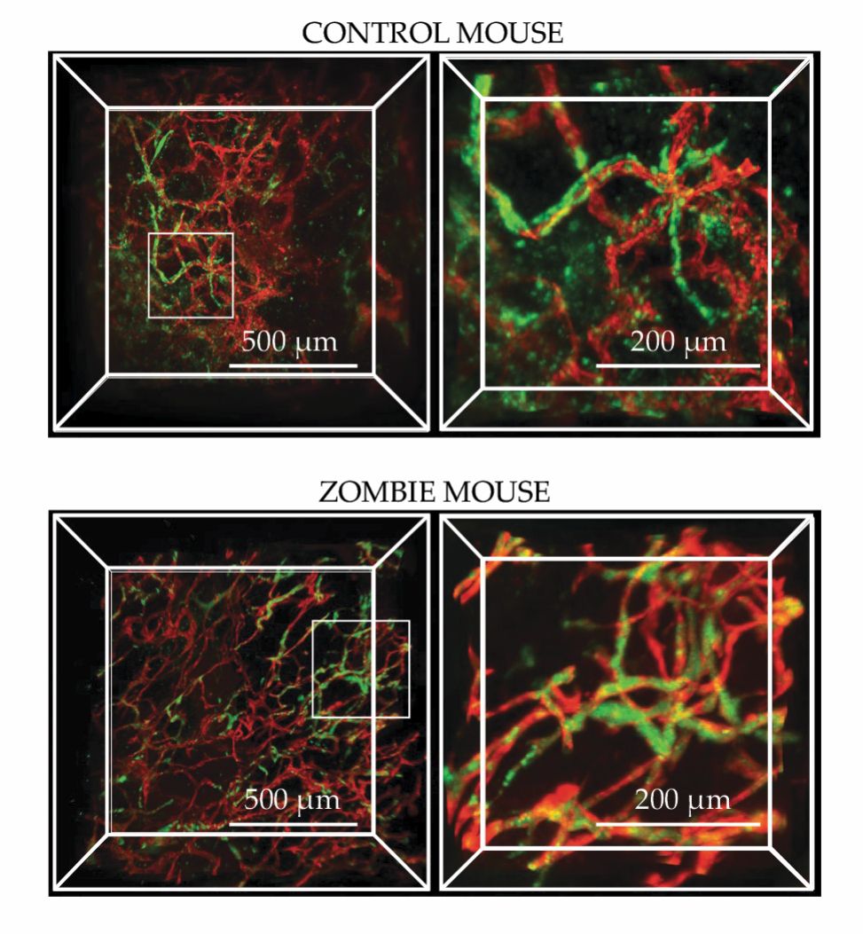
Analysis of the Zombie experiment implied that for 50 nm gold nanoparticles, passive inter-endothelial transport accounts for just 3% of all nanoparticles entering the tumor. (Curiously, that fraction was greater—as high as 25%—for both larger and smaller nanoparticles, but it was always a minority.) The rough agreement between the experiment and simulation offers some confirmation of both methods. “That was a pleasant surprise,” said Sindhwani, “and it gave us confidence.”
All lines of the Toronto researchers’ reasoning point to active trans-endothelial pathways as the primary means by which nanoparticles are delivered to tumors. But there’s still work to be done. All their experiments so far used nanoparticles made of gold, which are easy to image but may not be representative of all nanomaterials. It’s possible that passive transport doesn’t work in exactly the same way in Zombie mice as in live ones. And the experiments don’t reveal anything about what the predominant active molecular pathways are, what biomolecules are involved in regulating them, or—crucially for nanomedicine—how they compare to similar pathways at work in healthy tissue. “While we’ve made a first step,” says Sindhwani, “we need a major collaborative undertaking to figure this out.”
References
1. S. Sindhwani et al., Nat. Mater. (2020), doi:https://doi.org/10.1038/s41563-019-0566-2 .
2. S. Wilhelm et al., Nat. Rev. Mater. 1, 16014 (2016). https://doi.org/10.1038/natrevmats.2016.14
3. S. E. McNeil, Nat. Rev. Mater. 1, 16073 (2016); https://doi.org/10.1038/natrevmats.2016.73
K. Bourzac, Proc. Natl. Acad. Sci. USA 113, 12600 (2016). https://doi.org/10.1073/pnas.1616895113
More about the authors
Johanna L. Miller, jmiller@aip.org


